. 2008 Feb 29;331(1-2):127-39.
doi: 10.1016/j.jim.2007.12.002. Epub 2008 Jan 9.

Development of a novel chemokine-mediated in vivo T cell recruitment assay

Affiliations

Development of a novel chemokine-mediated in vivo T cell recruitment assay

Gabriele S V Campanella et al. J Immunol Methods. .

Abstract

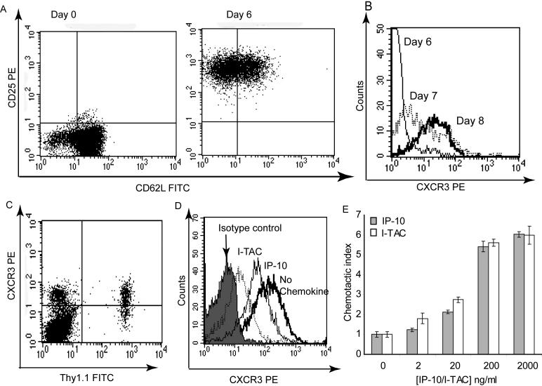
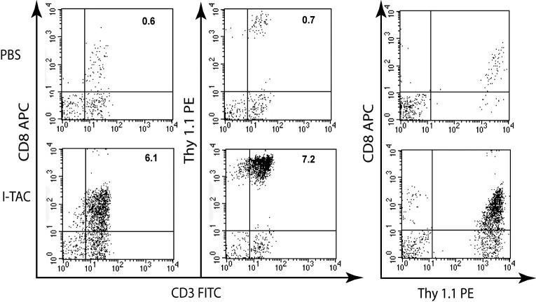
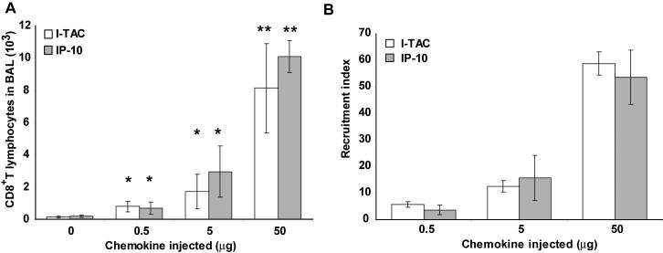
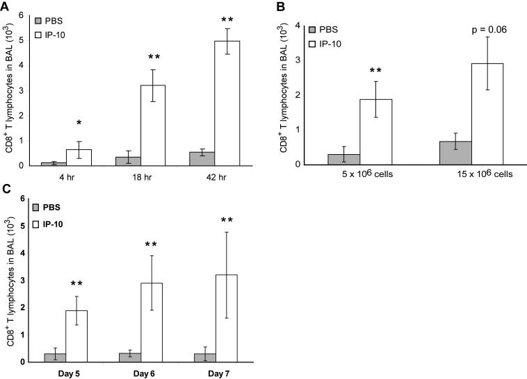
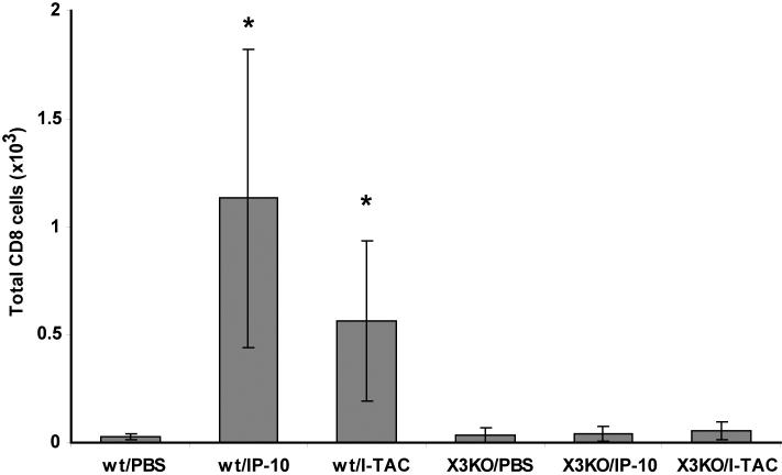
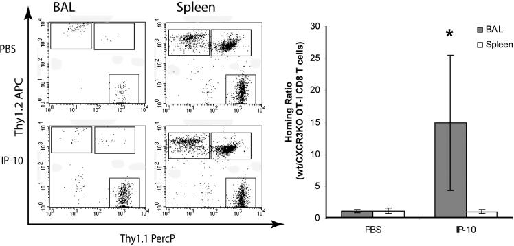
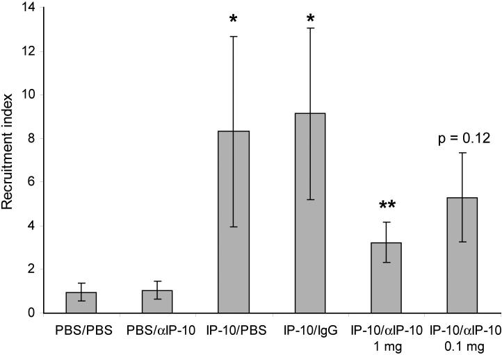
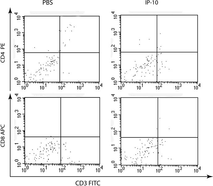
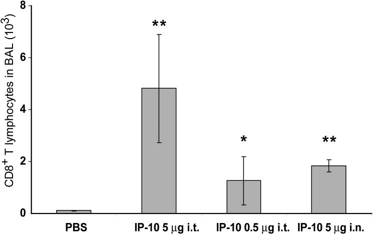
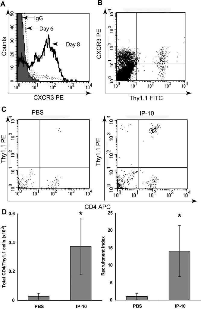
Trafficking of leukocytes to sites of inflammation is an important step in the establishment of an immune response. Chemokines are critical regulators of leukocyte trafficking and are widely studied molecules for their important role in disease and for their potential as new therapeutic targets. The ability of chemokines to induce leukocyte recruitment has been mainly measured by in vitro chemotaxis assays, which lack many components of the complex biological process of leukocyte migration and therefore provide incomplete information about chemokine function in vivo. In vivo assays to study the activity of chemokines to induce leukocyte recruitment have been difficult to establish. We describe here the development of a robust in vivo recruitment assay for CD8(+) and CD4(+) T lymphocytes induced by the CXCR3 ligands IP-10 (CXCL10) and I-TAC (CXCL11). For this assay, in vitro activated T lymphocytes were adoptively transferred into the peritoneum of naïve mice. Homing of these transferred T lymphocytes into the airways was measured following intratracheal instillation of chemokines. High recruitment indices were achieved that were dependent on chemokine concentration and CXCR3 expression on the transferred lymphocytes. Recruitment was also inhibited by antibodies to the chemokine. The assay models the natural condition of chemokine-mediated lymphocyte migration into the airways as chemokines are expressed in the airways during inflammation. The nature of this model allows flexibility to study wildtype and mutant chemokines and chemokine receptors and the ability to evaluate chemokine antagonists and antibodies in vivo. This assay will therefore help elucidate a deeper understanding of the chemokine system in vivo.


Figures

Figure 1
Figure 1. Characterization of in vitro activated CD8+ T lymphocytes
A) Expression of activation markers CD62L and CD25 on Day 0 and 6 of culture, as determined by Flow cytometry. B) Expression of CXCR3 on different days of in vitro culture. C) Expression of CXCR3 on Thy1.1+ T lymphocytes in the spleen three days after adoptive transfer of cells. D) CXCR3 internalization induced by 10 nM I-TAC or IP-10 E) Chemotaxis induced by I-TAC and IP-10. Each bar presents mean chemotactic index +/− STD. Experiments in D and E were performed with activated CD8+ T lymphocytes after eight days in culture.
Figure 2
Figure 2. Flow cytometric analysis of CD8+ T lymphocyte recruitment into the airways induced by I-TAC
I-TAC (5 μg) was injected intratracheally after adoptive transfer of activated CD8+ T lymphocytes 48 hr prior. The BAL was harvested 18 hr later, and CD8+ T lymphocytes were analyzed by flow cytometry after gating on the lymphocyte subpopulation. The percentages of CD8+/CD3+ T cells of total events are shown in the upper right corners.
Figure 3
Figure 3. Dose response of I-TAC and IP-10 induced recruitment of activated CD8+ T lymphocytes in vivo
PBS, I-TAC or IP-10 was injected intratracheally at the indicated amount. The BAL was harvested 18 hr later, and CD8+ T lymphocytes were analyzed by flow cytometry. A) Total CD8+ T lymphocytes in BAL. B) Recruitment index was calculated in comparison to intratracheal injection of PBS. * p < 0.05, ** p < 0.001 compared to PBS injection.
Figure 4
Figure 4. Variation of assay parameters
A) Timing of BAL: PBS or IP-10 (5 μg) was injected intratracheally after adoptive transfer of activated CD8+ T lymphocytes 48 hr prior. The BAL was harvested at the indicated time points, and CD8+ T lymphocytes were analyzed by flow cytometry. B) Number of CD8+ T lymphocytes adoptively transferred. PBS or IP-10 (5 μg) was injected intratracheally after adoptive transfer of activated CD8+ T lymphocytes as indicated 48 hr prior. The BAL was harvested after 18 hr, and CD8+ T lymphocytes were analyzed by flow cytometry. 2 mice per group were used for the 15×106 cells groups. C) Timing of in vitro culture: CD8+ T lymphocytes were activated in vitro for the indicated number of days and then adoptively transferred into mice. PBS or IP-10 (5μg) was injected intratracheally 48 hr later. The BAL was harvested after 18 hr, and CD8+ T lymphocytes were analyzed by flow cytometry. * p < 0.05, ** p < 0.01 each compared to PBS injection under the same conditions.
Figure 5
Figure 5. Transfer of wt or CXCR deficient activated CD8+ T cells
Activated wt or CXCR3 KO OT-I cells were adoptively transferred into mice. Forty-eight hours later, PBS, 5 μg I-TAC or IP-10 was injected intratracheally. The BAL was harvested 18 hr later, and CD8+ T lymphocytes were analyzed by flow cytometry. * p < 0.05 compared to PBS injection.
Figure 6
Figure 6. Co-transfer of wt and CXCR3 KO activated CD8+ T cells
Activated wt (Thy1.1) and CXCR3 KO (Thy1.2) OT-I cells (5×10⁶ each) were adoptively transferred into the same Thy1.1×Thy1.2 mice. Forty-eight hours later, PBS or 5 μg IP-10 was injected intratracheally. The BAL and spleen were harvested 18 hr later, and Thy1.1 and Thy1.2 single positive cells were analyzed by flow cytometry as shown after gating on CD3+/CD8+ T lymphocytes. The homing ratio was calculated as wt/CXCR3 KO OT-I CD8+ T lymphocytes. * p < 0.05 compared to PBS injection.
Figure 7
Figure 7. Inhibition of lymphocyte recruitment by antibody against IP-10
Anti-IP-10 or control antibody was injected i.p. into mice as indicated four hours before adoptive transfer of CD8+ T lymphocytes, and two hours after intratracheal injection of PBS or IP-10 (5 μg). The rest of the assay was performed as described for figure 2. * p < 0.01 compared to PBS injection, ** p < 0.02 compared to IP-10 instillation (with or without control antibody).
Figure 8
Figure 8. Recruitment of lymphocytes without adoptive cell transfer
PBS or IP-10 (5μg) was injected intratracheally into mice. The BAL was harvested after 18 hr, and CD8+ and CD4+ T cells were analyzed by flow cytometry as indicated.
Figure 9
Figure 9. Intratracheal or intranasal instillation of chemokine
IP-10 was injected intratracheally or intranasally into mice as indicated 48 hours after adoptive transfer of activated CD8+ T lymphocytes. The BAL was harvested 18 hr later and analyzed for CD8+ T lymphocytes by flow cytometry. * p < 0.05, ** p < 0.005 compared to PBS injection.
Figure 10
Figure 10. Adoptive transfer of activated CD4+ T lymphocytes
A) CXCR3 expression of CD4+ T lymphocytes after six or eight days in culture. B) CXCR3 expression of Thy1.1+ CD4+ T lymphocytes in the spleen three days after adoptive transfer. C) PBS or IP-10 (5 μg) was injected intratracheally into mice as indicated 48 hours after adoptive transfer of activated CD4+ T lymphocytes. The BAL was harvested 18 hr later and analyzed for Thy1.1/CD4+ T lymphocytes by flow cytometry. D) Quantitative representation of experiment performed in C). Recruitment indices were calculated in comparison to intratracheal injection of PBS. * p < 0.05 compared to PBS injection.

References

    1. Balashov KE, Rottman JB, Weiner HL, Hancock WW. CCR5(+) and CXCR3(+) T cells are increased in multiple sclerosis and their ligands MIP-1alpha and IP-10 are expressed in demyelinating brain lesions. Proc Natl Acad Sci U S A. 1999;96:6873–8. - PMC - PubMed
    1. Butcher EC. Leukocyte-endothelial cell recognition: three (or more) steps to specificity and diversity. Cell. 1991;67:1033–6. - PubMed
    1. Campanella GS, Grimm J, Manice LA, Colvin RA, Medoff BD, Wojtkiewicz GR, Weissleder R, Luster AD. Oligomerization of CXCL10 is necessary for endothelial cell presentation and in vivo activity. J Immunol. 2006;177:6991–8. - PubMed
    1. Charo IF, Ransohoff RM. The many roles of chemokines and chemokine receptors in inflammation. N Engl J Med. 2006;354:610–21. - PubMed
    1. Colvin RA, Campanella GS, Sun J, Luster AD. Intracellular domains of CXCR3 that mediate CXCL9, CXCL10, and CXCL11 function. J Biol Chem. 2004;279:30219–27. - PubMed

Publication types

MeSH terms