. 2008 Feb 29;376(4):1168-81.
doi: 10.1016/j.jmb.2007.12.003. Epub 2007 Dec 8.

RSV capsid polymorphism correlates with polymerization efficiency and envelope glycoprotein content: implications that nucleation controls morphogenesis

Affiliations

RSV capsid polymorphism correlates with polymerization efficiency and envelope glycoprotein content: implications that nucleation controls morphogenesis

Carmen Butan et al. J Mol Biol. .

Abstract

We used cryo-electron tomography to visualize Rous sarcoma virus, the prototypic alpharetrovirus. Its polyprotein Gag assembles into spherical procapsids, concomitant with budding. In maturation, Gag is dissected into its matrix, capsid protein (CA), and nucleocapsid moieties. CA reassembles into cores housing the viral RNA and replication enzymes. Evidence suggests that a correctly formed core is essential for infectivity. The virions in our data set range from approximately 105 to approximately 175 nm in diameter. Their cores are highly polymorphic. We observe angular cores, including some that are distinctively "coffin-shaped" for which we propose a novel fullerene geometry; cores with continuous curvature including, rarely, fullerene cones; and tubular cores. Angular cores are the most voluminous and densely packed; tubes and some curved cores contain less material, suggesting incomplete packaging. From the tomograms, we measured the surface areas of cores and, hence, their contents of CA subunits. From the virion diameters, we estimated their original complements of Gag. We find that Rous sarcoma virus virions, like the human immunodeficiency virus, contain unassembled CA subunits and that the fraction of CA that is assembled correlates with core type; angular cores incorporate approximately 80% of the available subunits, and open-ended tubes, approximately 30%. The number of glycoprotein spikes is variable (approximately 0 to 118) and also correlates with core type; virions with angular cores average 82 spikes, whereas those with tubular cores average 14 spikes. These observations imply that initiation of CA assembly, in which interactions of spike endodomains with the Gag layer play a role, is a critical determinant of core morphology.


Figures

Fig. 1
Fig. 1
Central section through a denoised tomogram, showing RSV virions of different sizes with cores of various kinds. Bar = 100 nm.
Fig. 2
Fig. 2
(A) Serial sections through a virion with an angular “coffin-like” core; and (B) Segmented surface renderings of the virion, showing both the outer surface studded with glycoprotein spikes (top) and the virion interior (bottom), with the capsid (red), internal material (orange) and material between the capsid and envelope (gray). Some densities connecting the capsid to the inner surface of the envelope are visible (e.g. arrows in A, row 2, column 2; row 3 column 3). In addition to the structurally homogeneous majority of surface protrusions representing Env spikes (see, e.g. A, row 3, column 1), the envelope contains a few morphologically distinct, presumably host-derived, protrusions (e.g. arrowheads A, row 3, column 2 and row 4, column 1; blow-ups in corners). The sections are 7.8 nm apart and 3.9 nm thick. Bar (A) = 50 nm.
Fig. 3
Fig. 3
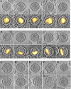
(A–I) Gallery of central sections through RSV virions with various kinds of cores. Bar (bottom right) = 50 nm. The panels in A and B are complemented with contrast-inverted versions of the same images containing surface renderings of the capsid surface (yellow). (A) Some virions with angular cores. Densities connecting the capsid to the envelope are marked with arrows (panels 2 and 5). (B) Some virions with “continuous curvature” cores. On average, these cores are smaller than angular cores and less densely packed. The core in panel 4 and perhaps also that in column 1 is a fullerene core (white asterisks). (C–E) Some virions with tubular cores, where the tubes may be open at both ends (C), at one end (D), or at neither end (E). Tubular cores tend not to be densely packed. (F–I) Virions with unusual, minority classes of cores. (F) A continuous curvature core appears to have been pressed tightly into the envelope, distorting its usually round shape. (G) Fullerene cone core, relatively densely packed and with a partial second layer capsid overlaid. (H) Virion lacking a well formed core. (I) Virion with two cores.
Fig. 4
Fig. 4
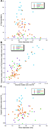
(A) Plot of number of spikes versus virion diameter. (B) Capsid internal volume expressed as a fraction of virion internal volume plotted versus virion diameter. (C) Plot of capsid surface area versus virion diameter. Each data point represents a virion, color-coded according to core type. One outlier with a larger diameter of 176 nm is not included.
Fig. 5
Fig. 5
Plot of number of assembled CA molecules assembled in a core versus the number of Gag molecules initially assembled into the provirion. If assembly were to be 100% efficient, these numbers would be equal, i.e. the data points would lie on the central diagonal. The difference between these numbers, (#Gag - #CA), represents the amount of unassembled CA in that virion. The arrowed lines at top give the estimated partition between assembled and unassembled CA for that virion. As in Fig. 4, each data point represents a virion, color-coded by core type. The colored dashed lines through the origin represent the average assembly efficiency for each core type. These data show that assembly efficiency varies systematically according to core type.
Fig. 6
Fig. 6
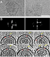
(A) Cryo-electron micrographs of isolated (incomplete) RSV capsids showing peripheral serrations with spacings of 4.1 nm (left) and 4.7 nm (right). (B) Diffraction patterns of the bracketed portions of (A), with the corresponding reflections marked (arrow). We infer that the serrations represent harmonics of a hexagonal lattice with 9.5 nm repeat (Results). The resolution (Table I) is insufficient to detect these spacings in intraviral capsids and the contrast of the 9.5 nm repeat is too low to be detected at prevailing noise levels. (C) Examples of tomographic sections through virions in which core-envelope linkers are visible (red arrows). Some of them underlie spike densities (yellow arrows). Bars (A, C) = 50 nm.
Fig. 7
Fig. 7
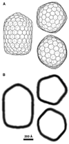
(A) Honeycomb model of an RSV capsid with the geometry of a fullerene coffin, shown in side view, top view and bottom view. This coffin has parameters (T1=16; T2= 9,13; Nr = 6). The cap (top) is that of a T=16 icosahedron; whereas the bottom has a (4+2) configuration of 5-fold vertices and pseudo-6-fold symmetry, corresponding to a mixture of T=9 and T=13 configurations. (B) Simulated density cross-sections, longitudinal and through the mid-section, at both ends.
Fig. 8
Fig. 8
Hypothetical model of proteolysis-induced maturation of the RSV provirion, including initiation of capsid assembly and genome packaging. (A) In the provirion, Gag polyproteins contact the host cell membrane where budding takes place (top) via their MA domains. Some may also interact with the less numerous Env spikes (bottom). (B) The protease, activated by being released from the polyprotein, processes the Gag molecules at their inter-domain cleavage sites. MA remains membrane-associated. The other domains enter soluble (or at least, disordered) pools in the virion interior (at right). (C) The putative initiation complex, containing of Env, Gag, and the viral RNA. The Gag shown here has lost its PR moiety but there is no reason to favor that model over unprocessed Gag. The NC domain binds RNA (and may already have done so in the provirion) to initiate packaging. The CA domains, possibly in a conformationally altered state or with other cofactor(s) bound, initiates assembly of CA subunits, leading to formation of a closed angular or curved capsid. The MA domains line the membrane. Absent an appropriate Env-Gag complex, polymerization of CA is eventually nucleated by a different mechanism with a higher critical concentration of CA subunits, producing a tubular capsid with low probability of successful packaging. The numbers of Env and Gag molecules in the putative initiation complex remain undefined. Alternatively, the nucleating principle may be Gag-Pol.

References

    1. Vogt VM. Retroviral Virions and Genomes. In: Coffin JM, Hughes SH, Varmus H, editors. Retroviruses. Woodbury, NY: Cold Spring Harbor Laboratory; 1997. pp. 27–70. - PubMed
    1. D'Souza V, Summers MF. How retroviruses select their genomes. Nat. Rev. Microbiol. 2005;3:643–655. - PubMed
    1. Steven AC, Heymann JB, Cheng N, Trus BL, Conway JF. Virus maturation: dynamics and mechanism of a stabilizing structural transition that leads to infectivity. Curr. Opin. Struct. Biol. 2005;15:227–236. - PMC - PubMed
    1. Briggs JA, Wilk T, Welker R, Kräusslich HG, Fuller SD. Structural organization of authentic, mature HIV-1 virions and cores. EMBO J. 2003;22:1707–1715. - PMC - PubMed
    1. Briggs JA, Watson BE, Gowen BE, Fuller SD. Cryoelectron microscopy of mouse mammary tumor virus. J. Virol. 2004;78:2606–2608. - PMC - PubMed

Publication types

LinkOut - more resources