

Functional abnormalities of the medial temporal lobe memory system in mild cognitive impairment and Alzheimer's disease: insights from functional MRI studies
- PMID: 18206188
- PMCID: PMC2760288
- DOI: 10.1016/j.neuropsychologia.2007.11.030
Functional abnormalities of the medial temporal lobe memory system in mild cognitive impairment and Alzheimer's disease: insights from functional MRI studies
Abstract
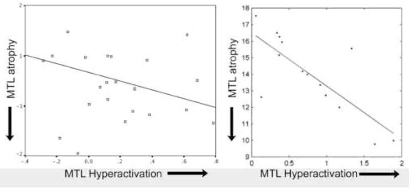
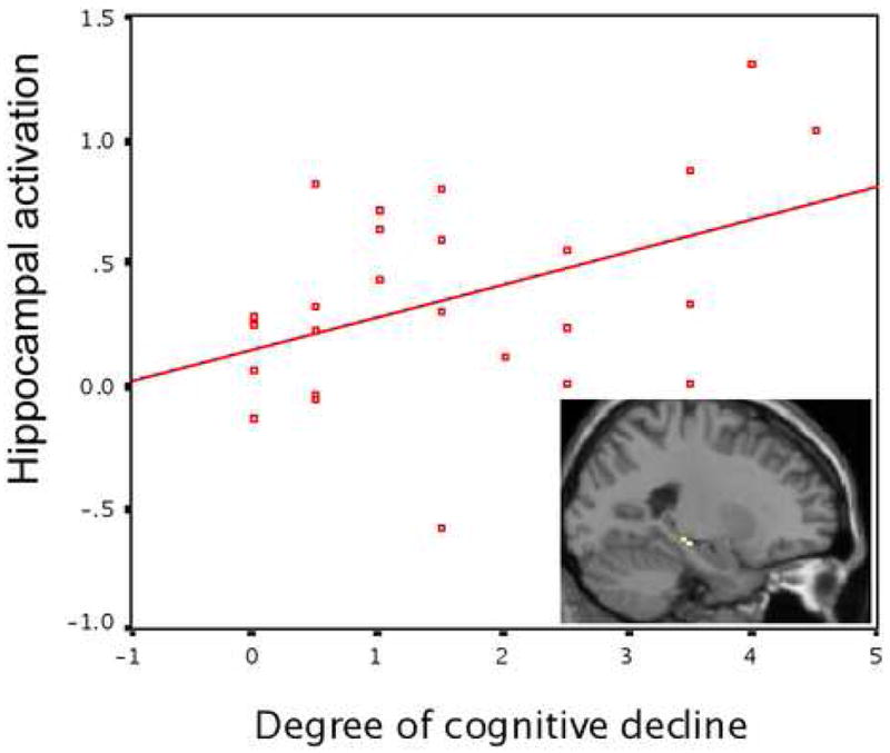
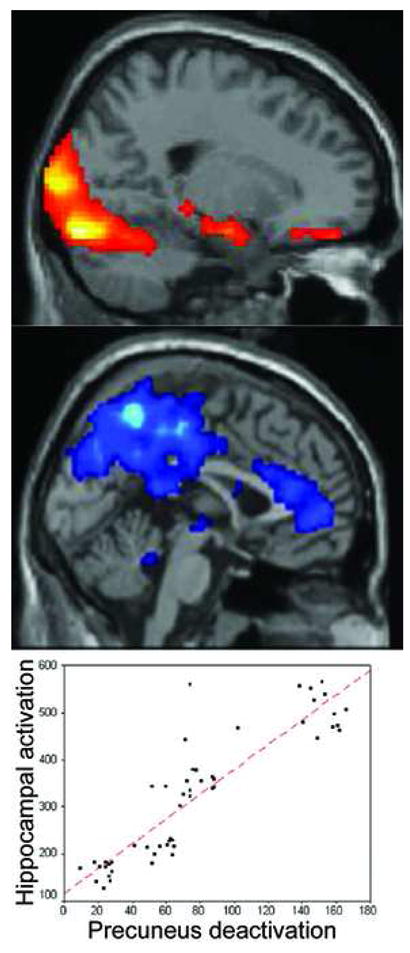
Functional MRI (fMRI) studies of mild cognitive impairment (MCI) and Alzheimer's disease (AD) have begun to reveal abnormalities in memory circuit function in humans suffering from memory disorders. Since the medial temporal lobe (MTL) memory system is a site of very early pathology in AD, a number of studies, reviewed here, have focused on this region of the brain. By the time individuals are diagnosed clinically with AD dementia, the substantial memory impairments appear to be associated with not only MTL atrophy but also hypoactivation during memory task performance. Prior to dementia, when individuals are beginning to manifest signs and symptoms of memory impairment, the hippocampal formation and other components of the MTL memory system exhibit substantial functional abnormalities during memory task performance. It appears that, early in the course of MCI when memory deficits and hippocampal atrophy are less prominent, there may be hyperactivation of MTL circuits, possibly representing inefficient compensatory activity. Later in the course of MCI, when considerable memory deficits are present, MTL regions are no longer able to activate during attempted learning, as is the case in AD dementia. Recent fMRI data in MCI and AD are beginning to reveal relationships between abnormalities of functional activity in the MTL memory system and in functionally connected brain regions, such as the precuneus. As this work continues to mature, it will likely contribute to our understanding of fundamental memory processes in the human brain and how these are perturbed in memory disorders. We hope these insights will translate into the incorporation of measures of task-related brain function into diagnostic assessment or therapeutic monitoring, such as for use in clinical trials.
Figures
References
-
- Arnold SE, Hyman BT, Flory J, Damasio AR, Van Hoesen GW. The topographical and neuroanatomical distribution of neurofibrillary tangles and neuritic plaques in the cerebral cortex of patients with Alzheimer’s disease. Cereb Cortex. 1991;1:103–116. - PubMed
-
- Backman L, Andersson JL, Nyberg L, Winblad B, Nordberg A, Almkvist O. Brain regions associated with episodic retrieval in normal aging and Alzheimer’s disease. Neurology. 1999;52:1861–1870. - PubMed
-
- Becker JT, Mintun MA, Aleva K, Wiseman MB, Nichols T, DeKosky ST. Compensatory reallocation of brain resources supporting verbal episodic memory in Alzheimer’s disease. Neurology. 1996;46:692–700. - PubMed
-
- Braak H, Braak E. Neuropathological stageing of Alzheimer-related changes. Acta Neuropathol (Berl) 1991;82:239–259. - PubMed
-
- Buckner RL, Snyder AZ, Sanders AL, Raichle ME, Morris JC. Functional brain imaging of young, nondemented, and demented older adults. J Cogn Neurosci. 2000;12(Suppl 2):24–34. - PubMed
Publication types
MeSH terms
Substances
Grants and funding
LinkOut - more resources
Full Text Sources
Medical
Research Materials
