Comparative Study
. 2008 Mar 1;315(1):72-88.
doi: 10.1016/j.ydbio.2007.12.008. Epub 2008 Jan 22.

Attenuation of retinal vascular development and neovascularization in PECAM-1-deficient mice

Affiliations
Comparative Study

Attenuation of retinal vascular development and neovascularization in PECAM-1-deficient mice

Terri A Dimaio et al. Dev Biol. .

Abstract

Platelet-endothelial cell adhesion molecule-1 (PECAM-1/CD31) is expressed on the surface of endothelial cells (EC) at high levels with important roles in angiogenesis and inflammation. However, the physiological role PECAM-1 plays during vascular development and angiogenesis remains largely unknown. Here we determined the role of PECAM-1 in the postnatal development of retinal vasculature and retinal neovascularization during oxygen-induced ischemic retinopathy (OIR) using PECAM-1-deficient (PECAM-1-/-) mice. A significant decrease in retinal vascular density was observed in PECAM-1-/- mice compared with PECAM-1+/+ mice. This was attributed to a decreased number of EC in the retinas of PECAM-1-/- mice. An increase in the rate of apoptosis was observed in retinal vessels of PECAM-1-/- mice, which was compensated, in part, by an increase in the rate of proliferation. However, the development and regression of hyaloid vasculature were not affected in the absence of PECAM-1. We did not observe a significant defect in astrocytes, the number of endothelial tip cell filopodias, and the rate of developing retinal vasculature progression in PECAM-1-/- mice. However, we observed aberrant organization of arterioles and venules, decreased secondary branching, and dilated vessels in retinal vasculature of PECAM-1-/- mice. In addition, retinal neovascularization was attenuated in PECAM-1-/- mice during OIR despite an expression of VEGF similar to that of PECAM-1+/+ mice. Mechanistically, these changes were associated with an increase in EphB4 and ephrin B2, and a decrease in eNOS, expression in retinal vasculature of PECAM-1-/- mice. These results suggest that PECAM-1 expression and its potential interactions with EphB4/ephrin B2 and eNOS are important for survival, migration, and functional organization of EC during retinal vascular development and angiogenesis.


Figures

Figure 1
Figure 1
Mouse retinal vasculature prepared by trypsin-digestion technique. Retinas were obtained from P42 wild type (A) or PECAM-1-/- mice (B). The arrows indicate EC, while arrowheads show the pericytes lining the retinal capillaries. Please note the decreased number of capillary loops as well as abnormal appearance of capillaries in the PECAM-1-/- retina. The quantitative assessment of the data is shown in Tables 1-3. Scale bar =20 μm (A and B).
Figure 2
Figure 2
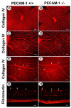
Collagen IV staining of the retinal wholemounts prepared from PECAM-1+/+ and PECAM-1-/- mice. Retinas were obtained from one-week- (A-D) or six-week-old (E-F) wild type (A, C, E) and PECAM-1-/- (B, D, F) mice and stained with an anti-collagen IV antibody as described in Experimental Procedures. Arrows indicate decreased number of capillary loops in retinas from PECAM-1-/- (D) compared with retinas from PECAM-1+/+ (C) mice. Please note decreased number of secondary branches off major arteries in retinas from PECAM-1-/- (F) compared with retinas from PECAM-1+/+ (E, arrows) mice. Arrowheads in E and F point to a major artery. Please note the larger diameter of arteries in retinas of PECAM-1-/- mice. G and H show fibronectin staining of frozen sections prepared from PECAM-1+/+ (G) and PECAM-1-/- (H) mice. Please note increased fibronectin staining in the retinal vessels of PECAM-1-/- mice which are dilated compared to PECAM-1+/+ mice (arrows). These experiments repeated three times with similar results. Scale bar (A and B = 200 μm; C and D = 100 μm; E and F = 20 μm; G and H = 50 μm).
Figure 3
Figure 3
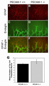
Astrocytic organization, formation of tip cells, and retinal vasculature expansion are not affected in the absence of PECAM-1. Wholemount retinas from PECAM-1+/+ (A, C, and E) and PECAM-1-/- (B, D, and F) P5 mice were stained with antibodies to GFAP (A and B) or endoglin (C and D). The merged images are shown in E and F. The quantitative assessment of tip cell’s filopodia is shown in G. Data in each bar are the mean number of filopodias counted in five retinas from five different mice. Please note similar organization and expansion of astrocytic template in PECAM-1+/+ and PECAM-1-/-. The endothelial tip cells similarly followed the astrocytic template without a significant difference in the number of tip cell filopodias. Other experiments were repeated twice with eyes from four different mice. Scale bar = 20 μm.
Figure 4
Figure 4
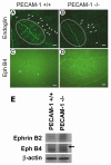
Aberrant organization of arterioles and venules and expression of EphB4 and Ephrin B2 in retinal vasculature of PECAM-1-/- mice. Wholemount retinas from P7 PECAM-1+/+ (A and C) and PECAM-1-/- (B and D) mice were stained with endoglin (A and B) or EphB4 (C and D). The exposure times were identical. The ovals in A and B mark an area where arterioles and venules are organizing. Please note the reduced density and aberrant organization of the arterioles and venules, and decreased secondary branching (arrowheads) in PECAM-1-/- mice. In addition, retinal vasculature of PECAM-1-/- mice showed significantly increased staining for EphB4. We were unable to detect Ephrin B2 in retinal wholemounts using antibodies from multiple sources. E shows a western blot of retinal extracts prepared from P7 PECAM-1+/+ and PECAM-1-/- mice and probed for EphB4 and Ephrin B2 as described in Methods. The blot was also probed with anti-β-actin to control for loading. These experiments were repeated three times with eyes from three different mice. Scale bar = 50 μm.
Figure 5
Figure 5
Increased rates of apoptosis in retinal vasculature of PECAM-1-/- mice. Wholemount trypsin-digested retinal vasculature were prepared from 2 weeks (A-D) or 3 weeks (E-H) old PECAM-1+/+ and PECAM-1-/- mice and processed for TUNEL staining as described in Methods. Panels A, B, E, and F show TUNEL staining (arrow heads). Panels C, D, G, and H show the merged TUNEL and Hoechst staining. The quantitative analysis of the data is shown in I. Data in each bar are the mean number of TUNEL positive cells per retina in five eyes of five mice (error bar indicate standard deviation). Please note a significant increase in the rate of apoptosis in PECAM-1-/- retinas compared to wild type mice (P< 0.01). These experiments were repeated twice with similar results. Scale bar = 100 μm.
Figure 6
Figure 6
Enhanced cell proliferation in retinal vasculature of PECAM-1-/- mice. Proliferating cells in P14 PECAM-1+/+ (A and C) and PECAM-1-/- (B and D) mice were labeled by BrdU and detected using an antibody to BrdU as described in Experimental Procedures. Collagen IV staining was used to visualize the vasculature. Quantitative assessment of cell proliferation on the retinal vessels is shown in D. Data in each bar are the mean number of BrdU+ cells per retina of five eyes of five mice (error bar indicate standard deviation). Please note that the number of proliferating cells are significantly higher in PECAM-1-/- retina compared to wild type (P<0.01). Scale bar ( A and B = 100 μm; C and D = 50 μm).
Figure 7
Figure 7
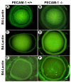
Assessment of hyaloid vasculature in wild type and PECAM-1-/- mice. The wholemount staining of hyaloid vasculature from wild type (A, C, and E ) and PECAM-1-/- (B, D, and F) mice at one week (A and B), two weeks (C and D), and three weeks (E and F) of age were prepared as described in Experimental Procedures. Please note similar vascularization and regression of vasculature in wild type and PECAM-1 -/- mice at all time points. Scale bar = 200 μm.
Figure 8
Figure 8
Quantitative assessments of the retinal nonperfused areas in response to hyperoxia. Wholemount retinas prepared from P12 wild type (A) or PECAM-1-/- (B) mice exposed to 5 days of hyperoxia were stained with antibody to collagen IV. Relative nonperfused areas of retinas were determined as described in Experimental Procedures (C). Data in each bar are the mean nonperfused area relative to whole area of each retina in five eyes of five mice (error bars indicate standard deviation). Please note that there is no significance difference in the degree of vessel obliteration in response to hyperoxia in wild type and PECAM-1 -/- mice (P = 0.73). Scal bar = 500 μm.
Figure 9
Figure 9
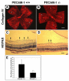
Quantitative assessment of neovascularization in P17 mice exposed to a cycle of hyperoxia and room air (OIR). A and B are collagen IV stained wholemount retinas, while C and D are HE/PAS stained eye sections prepared from P17 wild type and PECAM-1-/- mice, respectively. The arrows indicate growth of new vascular tufts which are significantly diminished in PECAM-1-/- mice. The number of vascular cell nuclei present on the vitreous side of the retina penetrating the inner limiting membrane was determined as described in experimental Procedures (E). Data in each bar are the mean number of vascular cell nuclei in five eyes of five mice (error bars indicate standard deviation). Please note that there is a significant decrease in the degree of retinal neovascularization in PECAM-1-/- mice compared with wild type mice (P <0.01). Scale bar (A and B = 500 μm; C and D = 50 μm).
Figure 10
Figure 10
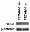
Assessment of VEGF levels in eyes from PECAM-1+/+ and PECAM-1 -/- mice. Eye extracts prepared from PECAM-1+/+ and PECAM-1-/- P15 mice (5 days of hyperoxia and 3 days of normoxia) were analyzed by SDS-PAGE and western blotting as described in Experimental Procedures. β-catenin was used as a loading control. These experiments were repeated twice with eyes from three different mice with similar results.
Figure 11
Figure 11
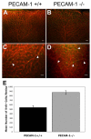
Reduced expression of eNOS in retinal vasculature of PECAM-1-/- mice. Retinal frozen sections prepared from P15 PECAM-1+/+ (A and C) and PECAM-1-/- (B and D) grown in room air (A and B) or exposed to OIR (5 days of hyperoxia and 3 days of room air) (C and D) were stained with an antibody to eNOS as described in Methods. All exposures were identical. Please note the PECAM-1+/+ retinal vasculature stained significantly stronger for eNOS, especially during OIR, when retinal neovascularization is occurring (arrows). These experiments were repeated three times with eyes from three different mice. Scale bar = 100 μm.

References

    1. Al-Shabrawey M, El-Remessy A, Gu X, Brooks SS, Hamed MS, Huang P, Caldwell RB. Normal vascular development in mice deficient in endothelial NO synthase: possible role of neuronal NO synthase. Mol Vis. 2003;9:549–58. - PubMed
    1. Albelda SM, Oliver PD, Romer LH, Buck CA. EndoCAM: a novel endothelial cell-cell adhesion molecule. J. Cell Biol. 1990;110:1227–1237. - PMC - PubMed
    1. Ando A, Yang A, Mori K, Yamada H, Yamada E, Takahashi K, Saikia J, Kim M, Melia M, Fishman M, Huang P, Campochiaro PA. Nitric oxide is proangiogenic in the retina and choroid. J Cell Physiol. 2002;191:116–24. - PubMed
    1. Bagi Z, Frangos JA, Yeh J-C, White CR, Kaley G, Koller A. PECAM-1 Mediates NO-Dependent Dilation of Arterioles to High Temporal Gradients of Shear Stress. Arterioscler Thromb Vasc Biol. 2005a;25:1590–1595. - PubMed
    1. Bagi Z, Frangos JA, Yeh JC, White CR, Kaley G, Koller A. PECAM-1 mediates NO-dependent dilation of arterioles to high temporal gradients of shear stress. Arterioscler Thromb Vasc Biol. 2005b;25:1590–5. - PubMed

Publication types

MeSH terms