. 2008 Feb 15;314(4):763-73.
doi: 10.1016/j.yexcr.2007.12.007. Epub 2008 Jan 22.

Dissection of the osteogenic effects of laminin-332 utilizing specific LG domains: LG3 induces osteogenic differentiation, but not mineralization

Affiliations

Dissection of the osteogenic effects of laminin-332 utilizing specific LG domains: LG3 induces osteogenic differentiation, but not mineralization

Robert F Klees et al. Exp Cell Res. .

Abstract

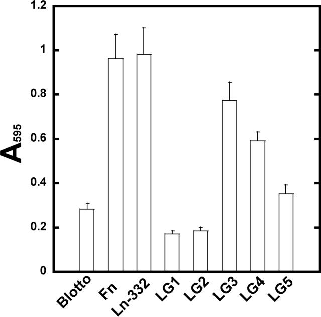
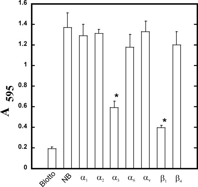
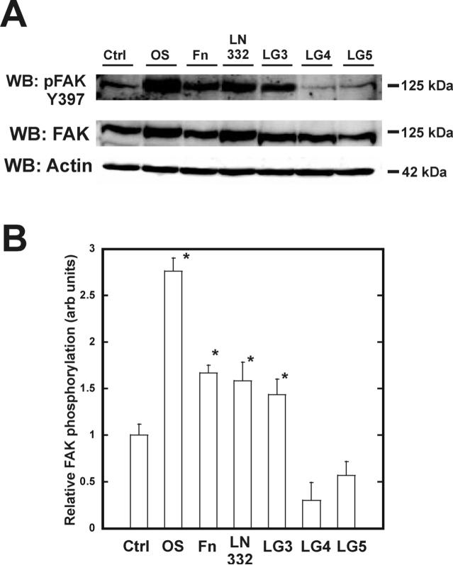
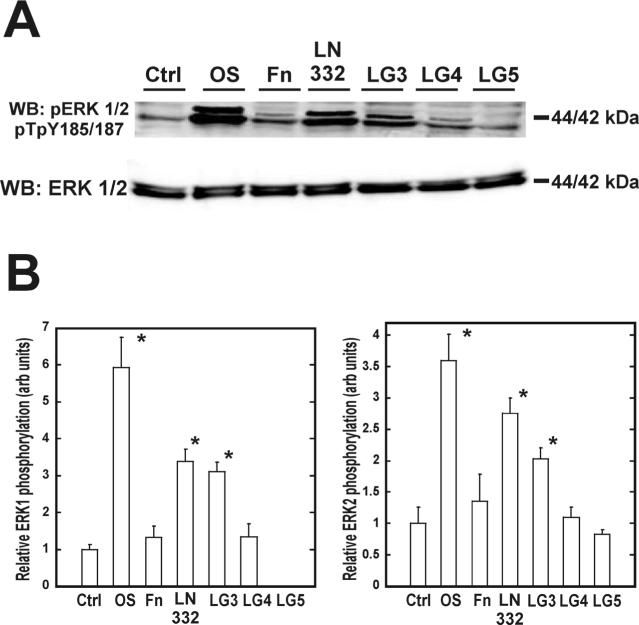
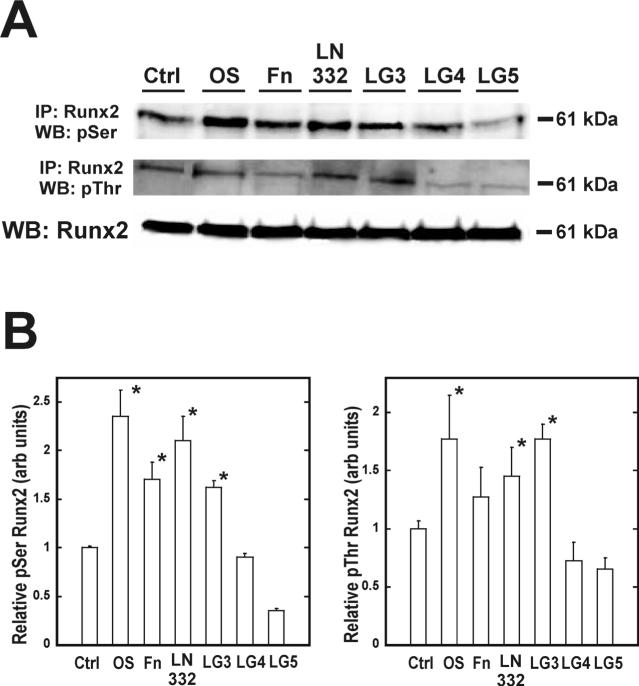
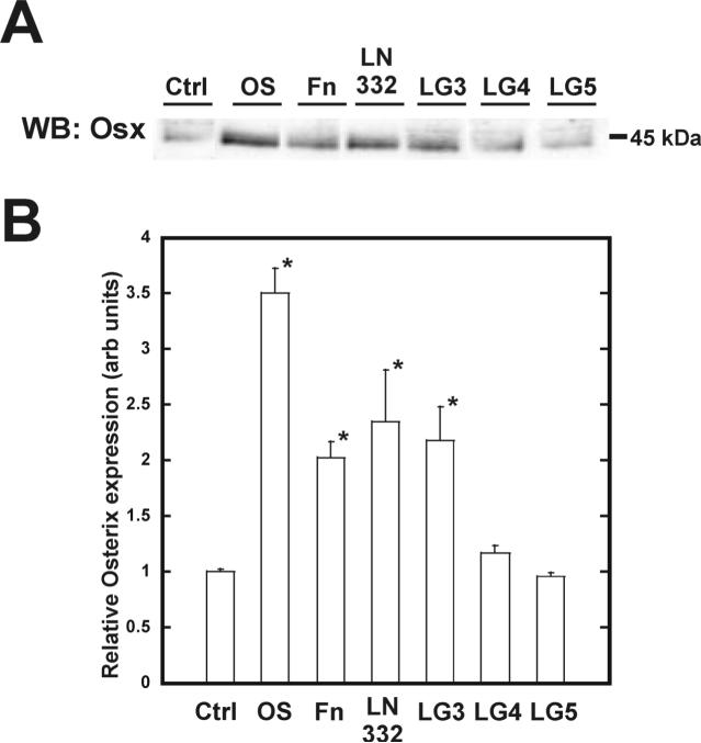
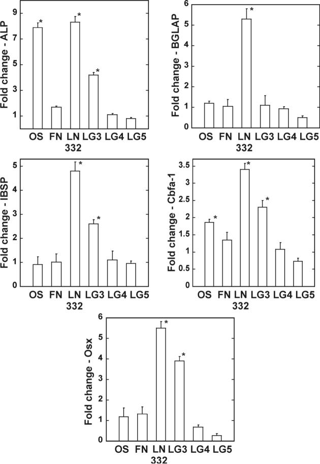
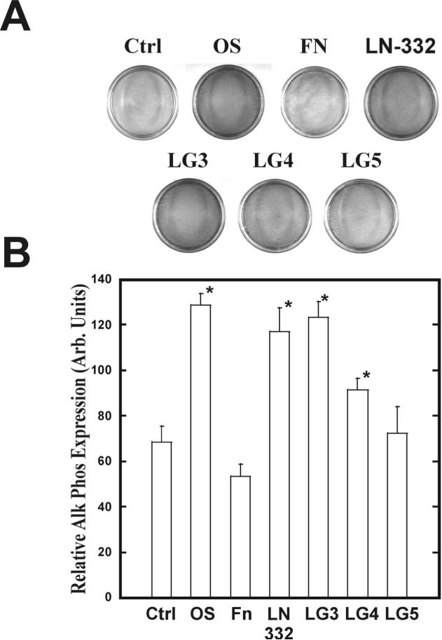
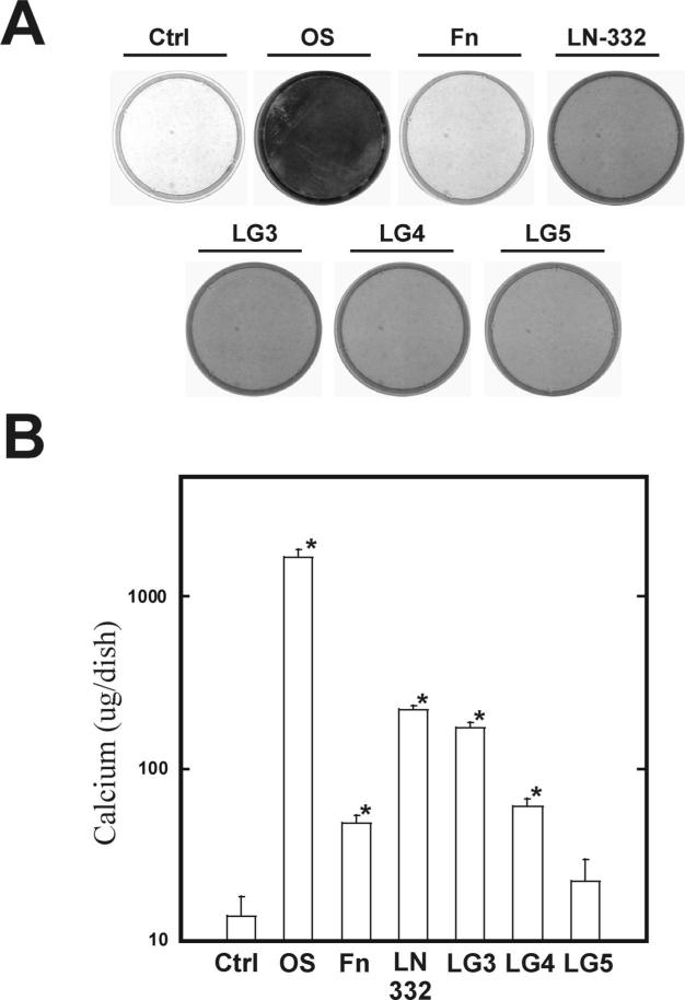
The overall mechanisms governing the role of laminins during osteogenic differentiation of human mesenchymal stem cells (hMSC) are poorly understood. We previously reported that laminin-332 induces an osteogenic phenotype in hMSC and does so through a focal adhesion kinase (FAK) and extracellular signal-related kinase (ERK) dependent pathway. We hypothesized that this is a result of integrin-ECM binding, and that it occurs via the known alpha3 LG3 integrin binding domain of laminin-332. To test this hypothesis we cultured hMSC on several different globular domains of laminin-332. hMSC adhered best to the LG3 domain, and this adhesion maximally activated FAK and ERK within 120 min. Prolonged culturing (8 or 16 days) of hMSC on LG3 led to activation of the osteogenic transcription factor Runx2 and expression of key osteogenic markers (osterix, bone sialoprotein 2, osteocalcin, alkaline phosphatase, extracellular calcium) in hMSC. LG3 domain binding did not increase matrix mineralization, demonstrating that the LG3 domain alone is not sufficient to induce complete osteogenic differentiation in vitro. We conclude that the LG3 domain mediates attachment of hMSC to laminin-332 and that this adhesion recapitulates most, but not all, of the osteogenic differentiation associated with laminin-5 binding to hMSC.


Figures

Figure 1
Figure 1. hMSC adhere best to the LG3 domain of laminin-332
hMSC were allowed to adhere to blotto (nfdm in PBST), 20 ug/ml FN, 10 ug/ml LN-332, 25 ug/ml LG1, LG2, LG4, LG5, 50 ug/ml LG3 for 2 hours. Non-adherent cells were removed by washing and adherent cells were stained with crystal violet, then solubilized in SDS and absorbance determined at 570nm.
Figure 2
Figure 2. Adhesion of hMSC to the LG3 domain of laminin-332 occurs through α3β1
hMSC were incubated with blocking antibodies to the indicated integrins and allowed to adhere to purified LG3 domain. Non-adherent cells were removed by washing and adherent cells were stained with crystal violet, then solubilized in SDS and absorbance determined at 570nm.
Figure 3
Figure 3. FAK Y397 phosphorylation occurs upon binding to the LG3 domain of laminin-332
hMSC were allowed to adhere for two hours on the indicated substrates and assayed for phosphorylated pY397 FAK (A). Densitometric measures of band intensities for pY397 FAK are shown in (B). Values were normalized to total protein and the two hour ctrl lane.
Figure 4
Figure 4. Adhesion of hMSC to the LG3 domain of laminin-332 induces phosphorylation of ERK 1/2
hMSC were allowed to adhere for two hours on the indicated substrates and assayed for phosphorylated pTpY185/187 ERK 1/2 (A). Densitometric measure of band intensities for ERK 1 and ERK 2 are shown in (B) and (C), respectively. ERK 1 and 2 bands were normalized to the intensity of ERK 1 and 2 in the two hour ctrl lane.
Figure 5
Figure 5. hMSC adhesion to LG3 domain increases phosphorylation of the master bone transcription factor, Runx2/Cbfa-1
Samples were incubated for 8 days, subsequently lysed, and Runx2/Cbfa-1 proteins immunoprecipitated with a Runx2/Cbfa-1 specific antibody. Immunoprecipitated proteins were separated by SDS-PAGE and blotted for phosphorylated serine and threonine, which is indicated by the 61 kDa band (A). Total Runx2/Cbfa-1 from each lysate was detected by western blot as a loading control. Densitometric measure of the ratio of band intensities for 61 kDa phosphoserine and phosphothreonine to total Runx2/Cbfa-1 are shown in (B) and (C), respectively.
Figure 6
Figure 6. The LG3 domain of laminin-332 amplifies the expression of the osteogenic transcription factor osterix
Samples were cultured as per figure 5 and blotted with an osterix specific antibody (A). Densitometric measure of band intensities for osterix are shown in (B).
Figure 7
Figure 7. Osteogenic gene expression is upregulated in hMSC grown on the LG3 domain of laminin-332
hMSC were plated as indicated and total mRNA isolated after day 16. Quantitative RT-PCR was performed on each sample using primers specific for the amplification of the indicated genes and normalized to GAPDH levels and gene expression from hMSC grown on tissue culture plastic.
Figure 8
Figure 8. Adhesion to LG domains increases osteogenic-specific marker expression of alkaline phosphatase in hMSC
hMSC were plated for 14 days as indicated and stained for alkaline phosphatase activity (A). Densitometry of plates shown in panel A (B).
Figure 9
Figure 9. The LG domains of laminin-332 increase the osteogenic-specific marker of calcified matrix in hMSC
Cells were cultured for 21 days on indicated substrates and stained for calcium using Alizarin Red (A). Colorimetric assessment of total calcium in hMSC (B). Cells were plated for 21 days as for panel A, then lysed and total calcium detected colorimetrically. Measurements were read at 570 nm.

Similar articles

Cited by

References

    1. Engvall E. Laminin variants: why, where and when? Kidney Int. 1993;43:2–6. - PubMed
    1. Malinda KM, Kleinman HK. The laminins. Int.J.Biochem.Cell Biol. 1996;28:957–959. - PubMed
    1. Nguyen NM, Senior RM. Laminin isoforms and lung development: all isoforms are not equal. Dev.Biol. 2006;294:271–279. - PubMed
    1. Stahl S, Weitzman S, Jones JC. The role of laminin-5 and its receptors in mammary epithelial cell branching morphogenesis. J.Cell Sci. 1997;110(Pt 1):55–63. - PubMed
    1. Kingsley K, Huff JL, Rust WL, Carroll K, Martinez AM, Fitchmun M, Plopper GE. ERK1/2 mediates PDGF-BB stimulated vascular smooth muscle cell proliferation and migration on laminin-5. Biochem.Biophys.Res.Commun. 2002;293:1000–1006. - PubMed

Publication types

MeSH terms