Comparative Study
. 2008 Jan 18;29(1):9-22.
doi: 10.1016/j.molcel.2007.11.031.

Nuclear FAK promotes cell proliferation and survival through FERM-enhanced p53 degradation

Affiliations
Comparative Study

Nuclear FAK promotes cell proliferation and survival through FERM-enhanced p53 degradation

Ssang-Taek Lim et al. Mol Cell. .

Abstract

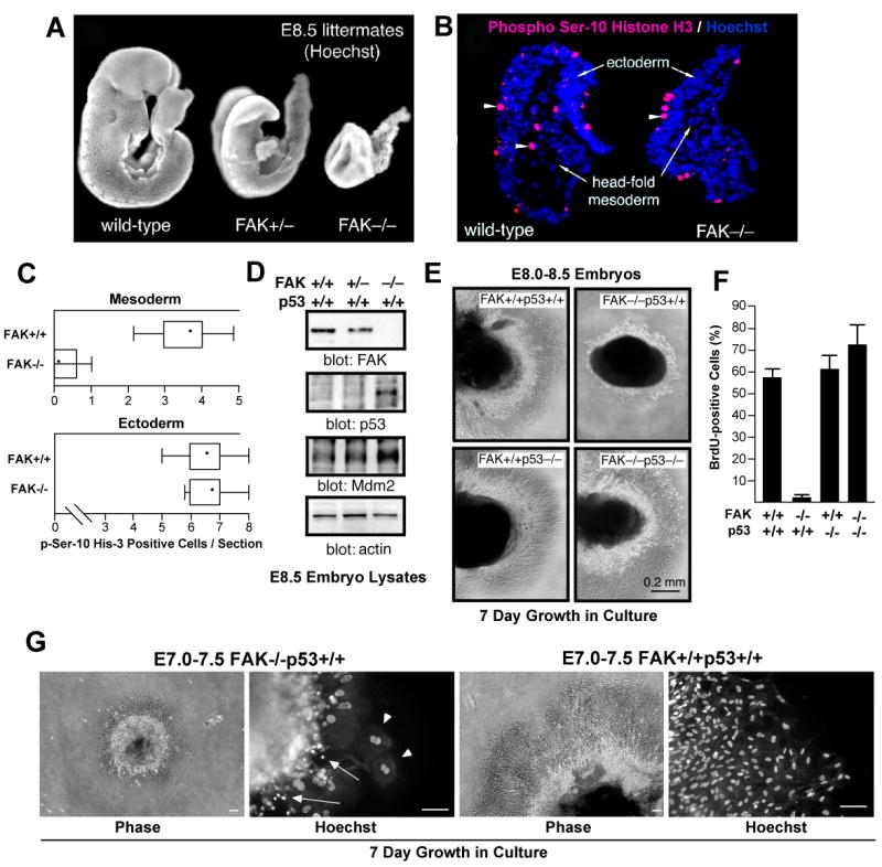
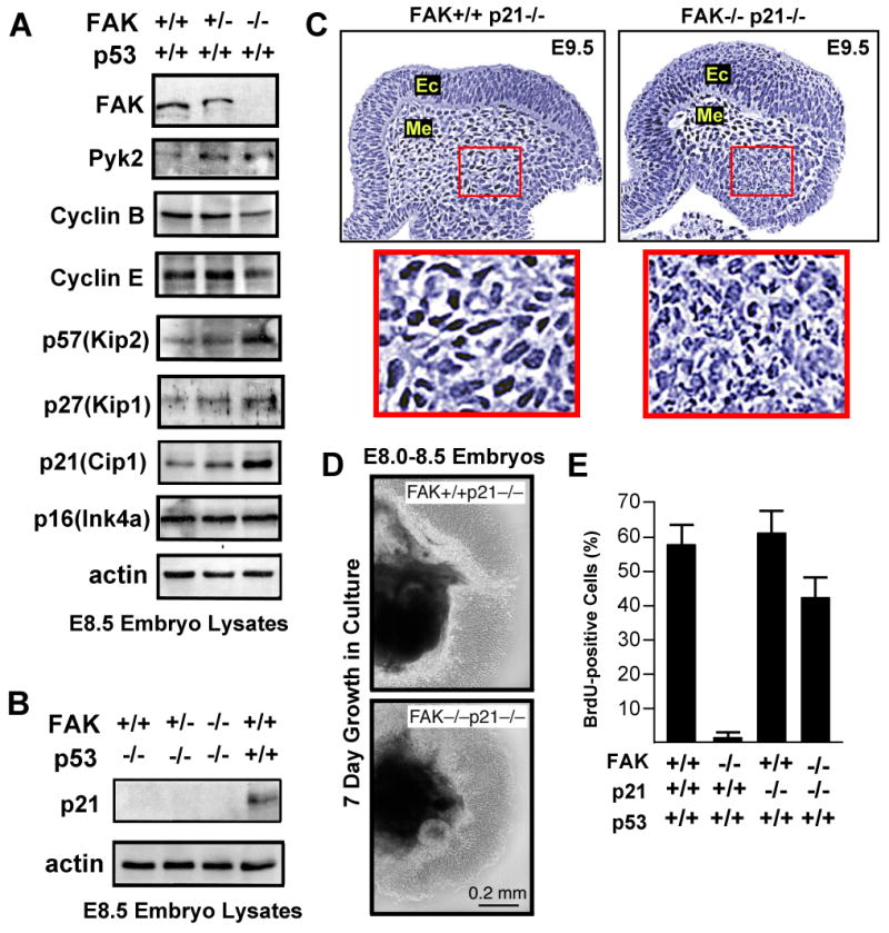
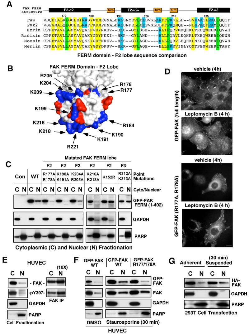
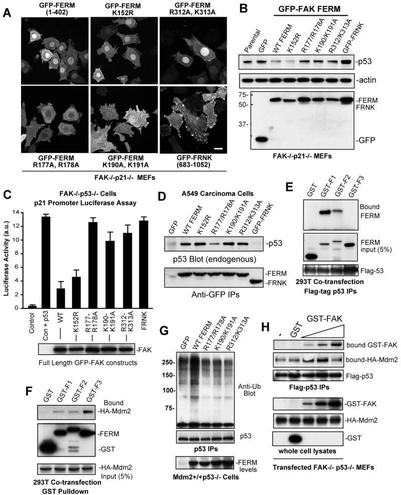
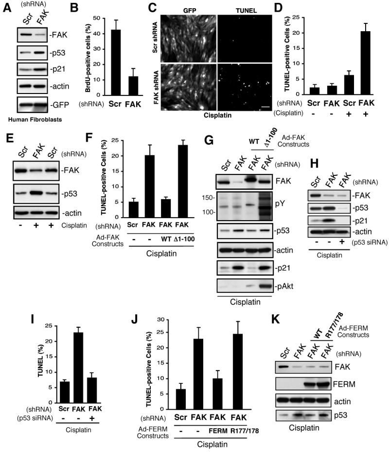
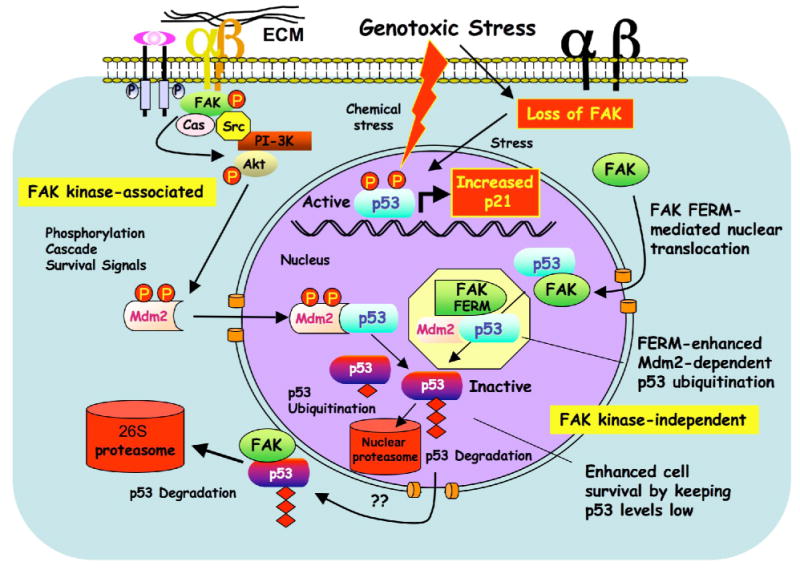
FAK is known as an integrin- and growth factor-associated tyrosine kinase promoting cell motility. Here we show that, during mouse development, FAK inactivation results in p53- and p21-dependent mesodermal cell growth arrest. Reconstitution of primary FAK-/-p21-/- fibroblasts revealed that FAK, in a kinase-independent manner, facilitates p53 turnover via enhanced Mdm2-dependent p53 ubiquitination. p53 inactivation by FAK required FAK FERM F1 lobe binding to p53, FERM F2 lobe-mediated nuclear localization, and FERM F3 lobe for connections to Mdm2 and proteasomal degradation. Staurosporine or loss of cell adhesion enhanced FERM-dependent FAK nuclear accumulation. In primary human cells, FAK knockdown raised p53-p21 levels and slowed cell proliferation but did not cause apoptosis. Notably, FAK knockdown plus cisplatin triggered p53-dependent cell apoptosis, which was rescued by either full-length FAK or FAK FERM re-expression. These studies define a scaffolding role for nuclear FAK in facilitating cell survival through enhanced p53 degradation under conditions of cellular stress.


Figures

Figure 1
Figure 1
FAK-/- embryo mesoderm cell proliferation block is p53-dependent. (A) Hoechst DNA staining of wild-type (FAK+/+), FAK+/-, and FAK embryos at E8.5. (B) Mesoderm region of FAK-/- embryos lack mitotic cells. Saggital head-fold sections of E8.0-8.5 FAK+/+ and FAK-/- littermate embryos stained with phosphoserine-10 Histone H3 (red, arrowheads) and Hoechst (blue). (C) Quantitation of phosphoserine-10 Histone H3 staining. Number of mitotic cells (per headfold region section) in the ectoderm and mesoderm of FAK-/- and FAK+/+ littermates plotted as Box-and-whisker diagrams: dot (mean), box (25 to 75 percentile), and bars (minimum and maximum value). (D) Immunoblotting of embryo lysates shows increased p53 and Mdm2 protein expression from E.8.5 FAK-/- compared to FAK+/+ or FAK+/- littermates. (E) p53 prevents FAK-/- embryo proliferation ex vivo. The indicated E8.0-8.5 embryos were dissected and cultured in Matrigel for 7 days. Phase contrast images of Matrigel-embedded embryos (dark) and surrounding cells growing out from the embryo mass. (F) Rescue of FAK-/- cell proliferation defects by p53 deletion. Anti-BrdU staining (green) and Hoechst (blue) labeling of dissociated cells from embryo explants cells at 40X. Percentage of BrdU-positive cells for the indicated genotype. Data are mean +/- SEM from 3 independent experiments. (G) E7.0-7.5 time of embryo extraction does not alter FAK-/-p53+/+ proliferation defects ex vivo. Phase images of embryos in Matrigel culture for 7 days (scale bar is 100 μm). Hoechst staining shows cells with multiple (arrowheads) and fragmented (arrows) nuclei in FAK-/-p53+/+ but not FAK+/+p53+/+ cultures.
Figure 2
Figure 2
p53-mediated proliferation block of FAK-/- cells is p21-dependent. (A) Increased expression of p53, Pyk2, or cyclin-dependent kinase inhibitors and decreased expression of cyclins in FAK-/- compared to FAK+/+ littermates as determined by immunoblotting of E.8.5 embryo protein lysates. (B) Lack of detectable p21 expression in lysates from FAK-/- E8.5 embryos on a p53-/- background. (C) FAK-/-p21-/- embryos exhibit lethality at E9.5. H&E staining of saggital headfold sections. Ec=ectoderm and Me=mesoderm. Inset, fragmented nuclei observed in mesoderm region of FAK-/-p21-/- but not FAK+/+p21-/- embryos. (D) E8.0-8.5 FAK-/-p21-/- embryo cells proliferate equally to FAK+/+p21-/- cells in 7 day Matrigel culture ex vivo. Phase contrast images of embryo mass (dark) and surrounding cells. (E) p21 inactivation promotes FAK-/- embryo cell proliferation as determined by the percentage of BrdU-positive cells counted for the indicated genotype. Data are mean +/- SEM from 3 independent experiments.
Figure 3
Figure 3
FAK FERM inhibits p53 activity through enhanced Mdm2-dependent ubiquitination and proteasomal degradation. (A) Epitope-tagged FAK construct schematic. Indicated is the band 4.1, ezrin, radixin, moesin (FERM) and central kinase domains. Filled regions indicate proline-rich motifs. Translation of FAK Δ1-100 starts at Met-101, Myc-tagged FERM encompasses residues 1-402, mutation of Y397 phosphorylation (F397), kinase-dead (KD, R454), Pro-null (Pro 712, 713, 872, 873, 876, and 877 mutated to Ala), and FRNK residues 691-1052 are indicated. (B) FAK FERM but not FAK kinase activity is required for the reduction of steady state p53 levels in FAK-/-p21-/- fibroblasts. Cells were transduced with Ad-Tet transactivator (TA, Mock), or Ad-TA plus the indicated Ad-FAK, Ad-FERM, or Ad-FRNK constructs. After 48 h, lysates were blotted with the indicated antibodies, p53 levels were quantified by densitometry, and mean values +/- SD are expressed as percentage of control (Mock) from 2 experiments. (C) Addition of MG132 (40 μM, 3h) prior to cell lysis prevents Ad-FAK and Ad-FERM-mediated p53 degradation in FAK-/-p21-/- fibroblasts. (D) FAK and FERM but not FRNK inhibit p53 activity as measured by a p21 promoter luciferase assay in FAK-/-p21-/- cells 48h after transfection. Values are means presented as percent of Mock control +/- SD from 3 experiments. (E) FAK and FERM but not FRNK promote enhanced p53 ubiquitination. HEK293 cells were co-transfected with flag-p53 and the indicated FAK constructs. MG132 was added 3h prior to lysis, p53 was isolated by IP, and analyzed by anti-ubiquitin and flag-tag blotting. (F) Mdm2 expression is required for FERM-enhanced p53 ubiquitination. Mdm2-/-p53-/- or Mdm2+/+p53-/- fibroblasts were transfected with flag-p53 and then transduced with Mock or Ad-FAK FERM. MG132 was added 3h prior to lysis, and p53 IPs were analyzed by anti-ubiquitin, anti-p53 (DO-1), and flag-tag blotting. Anti-Myc blotting was used to detect FAK FERM and anti-actin for loading control. Arrows indicate p53-shifted bands induced by FERM expression in Mdm2+/+ cells.
Figure 4
Figure 4
Determinants of FAK nuclear localization. (A) Structure-based alignment of FAK FERM F2 lobe residues (Lietha et al., 2007) with other FERM-containing proteins. Conserved basic residues within FAK and Pyk2 are highlighted in blue, total conserved FERM residues are highlighted in yellow and identical residues in green. (B) Localization of basic residue clusters on the surface of the FAK FERM F2 lobe. The FAK FERM domain (Lietha et al., 2007) F2 lobe was visualized using MacPyMOL. Basic residues (blue) are numbered according to the primary FAK sequence and acidic residues (red) are indicated. A putative nuclear targeting motif is comprised of residues at the tip of the F2 lobe (K190, K191, K216, K218, and R221). (C) FAK FERM domain analyzed by cellular fractionation. The indicated residues within GFP-FAK FERM (1-402) were mutated and constructs expressed in 293T cells. Cell lysates were separated into cytosolic (C) and nuclear (N) fractions, resolved by SDS-PAGE, and anti-GFP blotting was used to detect FAK FERM. Antibodies to gylceraldehyde-3-phosphate dehydrogenase (GAPDH) and poly ADP-ribose polymerase (PARP) were used to verify fractionation specificity, respectively. (D) Live cell imaging was used to follow GFP-FAK WT and GFP-FAK R177/178A distribution upon leptomycin B (10 ng/ml) or ethanol (vehicle) addition for 4 h. Scale bar is 10 μm. (E) FAK is partially nuclear-localized. HUVECs were separated into cytosolic and nuclear fractions, and blotted for FAK, GAPDH, and PARP. Ten-fold excess nuclear lysates was used to analyze FAK tyrosine phosphorylation by IP. (F) WT but not R177/178A FAK nuclear accumulation by HUVEC fractionation. HUVECs were treated with 1 μM staurosporine for 30 min, lysates separated into cytosolic or nuclear fractions, and immunoblotted with antibodies to GFP, FAK, GAPDH, and PARP. (G) 293T cells were transfected with HA-FAK and then fractionated into cytosolic and nuclear fractions under adherent and suspended conditions. Lysates blotted with anti-HA, GAPDH, and PARP.
Figure 5
Figure 5
Separate FAK FERM lobes mediate p53 binding, nuclear localization, and Mdm2 association. (A) The indicated GFP-FAK FERM (1-402) constructs or GFP-FRNK were stably-expressed in FAK-/-p21-/- (Pyk2 shRNA) fibroblasts and intracellular distribution visualized by confocal microscopy. Scale bar is 20 μm. (B) Steady-state p53 expression is reduced by FAK FERM expression in FAK-/-p21-/- (Pyk2 shRNA) fibroblasts as detected by p53, actin, and GFP bloting of lysates. (C) FERM domain mutations disrupt full length FAK inhibition of p53 transcriptional activity. FAK-/-p53-/- fibroblasts were transiently-transfected with a 2.4 kb p21 promoter luciferase construct (Vector, V) or in combination with p53 (Vec+p53) and the indicated FAK constructs. Luciferase activity is arbitrary units (a.u.). Values are means of 2 experiments +/- SD. Blotting verified equal FAK construct expression (below). (D) F2 FERM mutations can weaken p53 association. Ad-FAK FERM or FRNK constructs were expressed in A549 cells and association with endogenous p53 analyzed by IP and blotting. (E) FAK FERM F1 lobe binds p53. 293T cells were co-transfected with flag-p53 and the indicated FAK F1, F2, or F3 FERM lobes as GST fusion proteins. Anti-GST blotting of Flag IPs was used to detect FERM lobe association with p53. (F) FAK FERM F3 lobe binds Mdm2. 293T cells were co-transfected with HA-Mdm2 and the indicated FAK F1, F2, or F3 FERM lobes as GST fusion proteins. Cells were treated with MG132 prior to cell lysis (40 μM, 3h), incubated with glutathione agarose, and anti-HA blotting detected bound Mdm2. (G) FAK FERM mutations disrupt FERM-enhanced p53 ubiquitination. Mdm2+/+p53-/- fibroblasts were transfected with flag-p53, transduced with the indicated Ad-FAK FERM constructs, and MG132 was added 3h prior to lysis. p53 IPs were analyzed by anti-ubiquitin, flag-tag, and GFP blotting. (H) Biphasic FAK effects on Mdm2-p53 complex formation. HA-Mdm2 and flag-p53 were transfected into FAK-/-p53-/- fibroblasts, MG132 was added 3h prior to lysis, and GST (500 ng) or increasing amounts of recombinant GST-FAK (10 ng-500 ng) were added prior to p53 isolation by IP. Bound Mdm2 and FAK within a p53 complex were detected by anti-HA and GST blotting.
Figure 6
Figure 6
FAK controls human diploid fibroblast proliferation and p53-dependent apoptosis. (A) Lysates from scrambled (Scr) and FAK shRNA infected cells after 72 h were analyzed by anti-FAK, p53, p21, actin, and GFP blotting. (B) FAK shRNA inhibits human fibroblast proliferation. Cells were infected with the indicated lentivirus for 72 h, BrdU was added for 16h in growth media, and cells stained with anti-BrdU antibody. Mean values +/-SD are percent of total GFP-positive cells. (C, D) FAK shRNA sensitizes human fibroblasts to cisplatin-stimulated apoptosis. Cells were infected with the indicated lentivirus for 48 h, cisplatin (20 μg/ml) was added for 48 h, cells were fixed, and then analyzed by TUNEL staining. (C) Representative images of GFP-expressing and TUNEL-stained fibroblasts. Scale bar is 200 μm. (D) Mean values +/- SD for cisplatin-stimulated apoptosis were obtained by counting three TUNEL-stained 10X fields of cells from two coverslips. Only GFP-positive cells were counted and the data represents two independent experiments. (E) Elevated p53 levels in cisplatin-treated FAK shRNA-expressing fibroblasts as treated as in panel D and analyzed by anti-FAK, p53, and actin blotting. (F, G) FERM domain integrity is required for rescue of cisplatin-stimulated apoptosis. Cells were infected with Scr or FAK shRNA lentivirus (48 h), transducted with Ad-FAK or Ad-FAK (Δ1-100), and after 24h, cisplatin (20 μg/ml, 48 h) was added prior to analysis by TUNEL staining. (F) Mean values +/- SD for cisplatin-stimulated apoptosis were obtained as described for panel D. (G) FAK but not Δ1-100 FAK reverses cisplatin-stimulated increases in p53 and p21 expression as determined by blotting. Δ1-100 FAK activates Akt as determined by phospho-specific blotting. (H, I) FAK shRNA-enhanced cisplatin-stimulated apoptosis is p53 dependent. Fibroblasts were transfected with p53 siRNA, transduced with Scr or FAK shRNA lentivirus, and treated with cisplatin (20 μg/ml, 48 h). (H) Blotting for FAK, p53, and p21 blotting show changes in protein expression with actin blotting as control. (I) Mean values +/- SD for cisplatin-stimulated apoptosis were obtained as for panel d. (J, K) FAK FERM domain rescue of cisplatin-stimulated apoptosis. Cells were transduced with Scr or FAK shRNA lentivirus (48 h), infected with Ad-Myc-FERM WT or Ad-Myc-FERM R177/R178 (24 h), and then treated with cisplatin (20 μg/ml, 48h). (J) Cells were analyzed fro TUNEL staining as in panel D. (K) Blotted for FAK, Myc tag (FERM), actin, and p53 shows that FERM mutation blocks p53 regulation.
Figure 7
Figure 7
Model of FAK FERM-mediated p53 turnover and cell survival. FAK can function with integrins and growth factor receptors to promote cell survival through signaling cascades such as Akt that can activate ubiquitin E3-ligases such as Mdm2 to maintain low p53 levels. This canonical survival pathway involves FAK kinase activity (left). Under reduced integrin adhesion or conditions of cellular stress, FAK leaves focal contacts sites. This increases the cytoplasmic pool of FAK and enhances FAK nuclear accumulation via FAK-FERM-mediated targeting. Nuclear FAK acts as a scaffold to stabilize a p53-Mdm2 complex, leading to p53 polyubiquitination, and subsequent p53 degradation by nuclear or cytoplasmic proteasomes. This regulatory connection between FAK and p53 is dependent on the FAK FERM domain, but does not require FAK kinase activity (right).

References

    1. Bao W, Thullberg M, Zhang H, Onischenko A, Stromblad S. Cell attachment to the extracellular matrix induces proteasomal degradation of p21(CIP1) via Cdc42/Rac1 signaling. Mol Cell Biol. 2002;22:4587–4597. - PMC - PubMed
    1. Braren R, Hu H, Kim YH, Beggs HE, Reichardt LF, Wang R. Endothelial FAK is essential for vascular network stability, cell survival, and lamellipodial formation. J Cell Biol. 2006;172:151–162. - PMC - PubMed
    1. Bryant P, Zheng Q, Pumiglia K. Focal adhesion kinase controls cellular levels of p27/Kip1 and p21/Cip1 through Skp2-dependent and -independent mechanisms. Mol Cell Biol. 2006;26:4201–4213. - PMC - PubMed
    1. Cohen LA, Guan JL. Residues within the first subdomain of the FERM-like domain in focal adhesion kinase are important in its regulation. J Biol Chem. 2005;280:8197–8207. - PubMed
    1. Cox BD, Natarajan M, Stettner MR, Gladson CL. New concepts regarding focal adhesion kinase promotion of cell migration and proliferation. J Cell Biochem. 2006;99:35–52. - PubMed

Publication types

MeSH terms