. 2008 Jan 18;29(1):102-11.
doi: 10.1016/j.molcel.2007.12.015.

The putative cancer stem cell marker USP22 is a subunit of the human SAGA complex required for activated transcription and cell-cycle progression

Affiliations

The putative cancer stem cell marker USP22 is a subunit of the human SAGA complex required for activated transcription and cell-cycle progression

Xiao-Yong Zhang et al. Mol Cell. .

Abstract

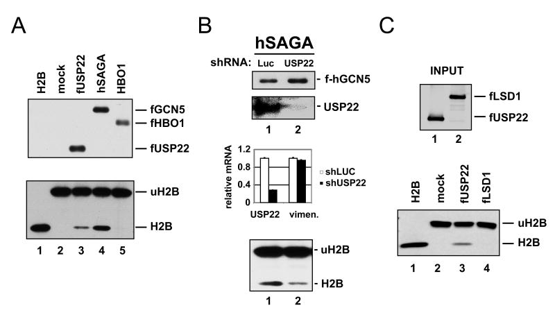
Polycomb genes encode critical regulators of both normal stem cells and cancer stem cells. A gene signature that includes Polycomb genes and additional genes coregulated with Polycomb genes was recently identified. The expression of this signature has been reported to identify tumors with the cancer stem cell phenotypes of aggressive growth, metastasis, and therapy resistance. Most members of this 11 gene signature encode proteins with well-defined roles in human cancer. However, the function of the signature member USP22 remains unknown. We report that USP22 is a previously uncharacterized subunit of the human SAGA transcriptional cofactor complex. Within SAGA, USP22 deubiquitylates histone H2B. Furthermore, USP22 is recruited to specific genes by activators such as the Myc oncoprotein, where it is required for transcription. In support of a functional role within the Polycomb/cancer stem cell signature, USP22 is required for appropriate progression through the cell cycle.


Figures

Figure 1
Figure 1. The cancer stem cell marker USP22 is a subunit of the hSAGA cofactor complex
(A) USP22 is a putative ubiquitin hydrolase containing a C-terminal peptidase domain of the C19 family. In addition, USP22 contains an amino terminal zinc finger motif. (B) A stable cell line expressing a FLAG-epitope tagged version of USP22 (fUSP22) was generated in the human non-small cell lung carcinoma line H1299. The FLAG-USP22 protein was affinity purified and co-purified proteins detected by silver staining. A mock purification was performed in parallel using extracts from parental cells. MS/MS identification of USP22 associated proteins was performed on bands excised from duplicate samples run in adjacent lanes and colloidal-stained. The polypeptides listed were identified in the MS/MS analysis and represent known components of the human SAGA complex. Peptide sequences from the recovered proteins are listed in Supplemental Table 1, with the total coverage ranging from 1-5%. Asterisks indicate known contaminants, including keratin and myosin. (C) The purified FLAG-USP22 complex was analyzed by western blotting for USP22 and the hSAGA subunits TRRAP and hGCN5. During affinity purification, four sequential eluates were harvested, as indicated. (D) Purified USP22 complex was assessed for HAT activity using an in vitro reaction containing core histones and radiolabeled acetyl-CoA (lower panel). Western blotting again confirmed the association of USP22 with the hSAGA subunits TRRAP and hGCN5, as well as the presence of FLAG-USP22 itself (upper panels). (E) Western blots confirmed that endogenous USP22 is associated with hSAGA. hSAGA was affinity purified from cells stably expressing FLAG-hGCN5. As a control, a cell line expressing FLAG-spRNAP was utilized. spRNAP and hGCN5 precipitation was confirmed by blotting for the FLAG epitope (top panel). Probing with anti-USP22 revealed the specific association of endogenous USP22 with the hSAGA complex (lower panel). (F) Endogenous hGCN5 and USP22 coprecipitate from nuclear extracts of human cells. Nuclear extracts from 293T cells were subjected to immunoprecipitation with an antibody recognizing endogenous USP22. Precipitates were resolved by SDS/PAGE and blotted for either USP22 (bottom panel) or hGCN5 (top panel). Control precipitates were conducted in parallel using nonimmune IgG, as indicated.
Figure 2
Figure 2. USP22 catalyzes the deubiquitylation of histone H2B in vitro
(A) FLAG-tagged USP22 or HBO1 were expressed in H1299 cells and affinity purified under non-denaturing conditions. In parallel, hSAGA was purified via FLAG-tagged hGCN5. The purification of USP22, hGCN5 and HBO1 was confirmed by western blotting for the FLAG epitope (upper panel). Purified USP22 complex, hSAGA and HBO1 complex were incubated in vitro with ubiquitylated H2B (uH2B). Reaction mixtures were then resolved by SDS/PAGE and developed with antisera against H2B (lower panel). In addition to deubiquitylation reactions that included the USP22 complex (lane 3), hSAGA (lane 4) and HBO1 (lane 5), a mock reaction was analyzed in parallel (lane 2). Purified non-ubiquitylated H2B was run as a migration standard (lane 1). (B) The contribution of USP22 to the deubiquitylation activity associated with hSAGA was assess by using shRNA to deplete USP22 from the FLAG-hGCN5 expressing cells. Control cells were infected with shRNA targeting luciferase (luc). Western blotting confirmed the purification of hGCN5 and the knockdown of endogenous USP22 within purified hSAGA (upper panels). Efficient USP22 depletion was also documented at the mRNA level using qRT-PCR (middle panel). hSAGA purified from control (luc) and USP22-depleted cells was subjected to an in vitro deubiquitylation assay with uH2B as in (A), (lower panel). Error bars represent standard deviation. (C) Recombinant USP22 was produced by baculovirus infection of insect cells and then purified via the FLAG epitope tag. The histone demethylase LSD1 was affinity purified in parallel, as a negative control. Purified proteins were detected by western blotting for the FLAG epitope (upper panel). The proteins were then subjected to an in vitro deubiquitylation assay as in A and B, using uH2B as a substrate, with H2B and uH2B detected by western blotting (lower panel).
Figure 3
Figure 3. USP22 is required for activator-driven transcription
(A) Normal diploid human fibroblasts were engineered to express a conditional allele of MYC that is activated by 4-OHT treatment. These cells were subjected to shRNA-mediated depletion of USP22 (shUSP22) or to control shRNA infection (shLUC). After MYC activation, mRNA levels of the indicated genes were determined by qRT-PCR. USP22 depletion efficiency was also determined by qRT-PCR (left panel). Fold induction for several MYC target genes was measured in the presence or absence of USP22 shRNA. These targets included cyclin D2 (CCND2), ODC, JPO1, CAD and MTA1. As a negative control, vimentin levels were quantified. All samples were normalized to ELF1a mRNA. (B) H1299 cells expressing a tet-regulated p53 allele were treated with USP22 or control shRNA (shLUC). p53 was induced for 8 or 24 hours by tetracycline treatment (1.0μg/ml), as indicated. mRNA levels for USP22 and the p53 target genes PUMA, p21 and PIG3 were quantitated, normalized and displayed as in (A). Error bars represent standard deviation.
Figure 4
Figure 4. Activator-dependent recruitment of USP22 to target gene loci
(A) H1299 cells were treated with shRNA directed against c-myc. MYC protein levels were assessed by western blotting (left panel), and qRT-PCR used to define levels of the MYC target genes CAD and MTA1 (right panel). (B) and (C) Within the CAD and MTA1 loci, binding of endogenous MYC and USP22 were evaluated by ChIP. The intron (solid line) and exon (dark boxes) structure of CAD and MTA1 are displayed. For both genes, documented MYC binding sites (labeled 5′) occur near the transcriptional start site (indicated by arrow). ChIP primer sets in the 3′ end of each locus were used to assess MYC and USP22 occupancy. As a control, nonimmune rabbit antibody (IgG) was used. The relative binding of USP22 and MYC to the CAD and MTA1 loci in the presence (white bars) or absence (black bars) of MYC shRNA is displayed. Error bars represent standard deviation.
Figure 5
Figure 5. USP22 is required for MYC-mediated transformation and for appropriate cell cycle progression
(A) After USP22 depletion, Tert-immortalized, p16 null human fibroblasts transformed by MYC, were analyzed for growth in soft agar. Luciferase shRNA-treated cells served as a control. Images show representative colony size and number at 10 days after USP22 knockdown (left panels). Colony number was also quantitiated by using microscopy to count all visible colonies, under low power in three 6 cm plates (right panel). This methodology resulted in scoring of all colonies containing > 15-20 cells. The soft agar assay was performed on three independent occasions using cells infected with separate batches of USP22 shRNA and the quantitation shown is a representative assay. Statistical analysis was performed on the colony number data from the USP22 knockdown and control groups (Student's t test), resulting in a P value of 0.001. Error bars represent standard deviation. (B) H1299 cells were infected with lentivirus expressing shRNA molecules directed against either USP22 (closed triangles) or luciferase (closed diamonds). After selection for infected cells, cells were plated in 6-well plates. Cell numbers were determined by direct counting of triplicate wells at each of the time points indicated. (C) H1299 cells from (B) were also stained with propidium iodide and cell cycle profile determined by flow cytometry.

Comment in

References

    1. Barlev NA, Liu L, Chehab NH, Mansfield K, Harris KG, Halazonetis TD, Berger SL. Acetylation of p53 activates transcription through recruitment of coactivators/histone acetyltransferases. Mol Cell. 2001;8:1243–1254. - PubMed
    1. Bello-Fernandez C, Packham G, Cleveland JL. The ornithine decarboxylase gene is a transcriptional target of c-Myc. Proc Natl Acad Sci U S A. 1993;90:7804–7808. - PMC - PubMed
    1. Ben-Saadon R, Zaaroor D, Ziv T, Ciechanover A. The polycomb protein Ring1B generates self atypical mixed ubiquitin chains required for its in vitro histone H2A ligase activity. Mol Cell. 2006;24:701–711. - PubMed
    1. Bhaumik SR, Green MR. SAGA is an essential in vivo target of the yeast acidic activator Gal4p. Genes Dev. 2001;15:1935–1945. - PMC - PubMed
    1. Blagosklonny MV. Target for cancer therapy: proliferating cells or stem cells. Leukemia. 2006;20:385–391. - PubMed

Publication types

MeSH terms