. 2008 Feb;38(2):190-200.
doi: 10.1016/j.ibmb.2007.10.008. Epub 2007 Nov 17.

Characterization of a juvenile hormone-regulated chymotrypsin-like serine protease gene in Aedes aegypti mosquito

Affiliations

Characterization of a juvenile hormone-regulated chymotrypsin-like serine protease gene in Aedes aegypti mosquito

Guowu Bian et al. Insect Biochem Mol Biol. 2008 Feb.

Abstract

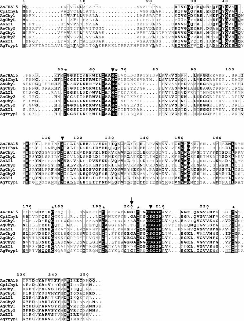
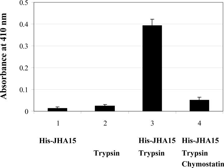
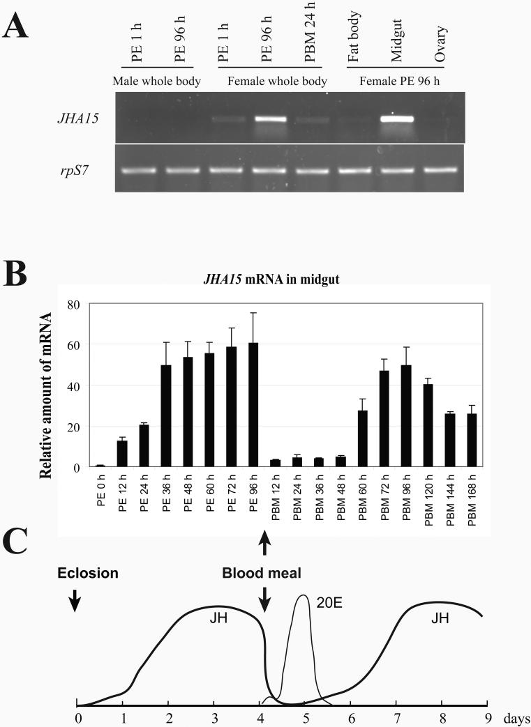
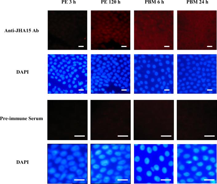
After female mosquitoes ingest blood from vertebrate hosts, exopeptidases and endopeptidases are required for digesting blood proteins in the midgut into amino acids, which female mosquitoes use to build yolk proteins. These proteases are not always present in the midgut, and their diverse expression patterns suggest that production of these enzymes is highly regulated in order to meet specific physiological demands at various stages. Here we report identification of a serine-type protease, JHA15, in the yellow fever mosquito Aedes aegypti. This protein shares high sequence homology with chymotrypsins, and indeed exhibits specific chymotrypsin enzymatic activity. The JHA15 gene is expressed primarily in the midgut of adult female mosquitoes. Our results indicate that its transcription is activated by juvenile hormone in the newly emerged female adults. Although its mRNA profile is similar to that of the early trypsin gene, we found that JHA15 proteins were readily detected in the midgut epithelium cells of both non-blood-fed and blood-fed mosquitoes. Analysis of polysomal RNA further substantiated that synthesis of JHA15 occurs before and shortly after blood feeding. Knocking down expression of JHA15 resulted in no evident phenotypic changes, implying that functional redundancy exists among those proteolytic enzymes.


Figures

Figure 1
Figure 1
Enrichment of JHA15 cDNAs by suppression subtractive hybridization. For forward subtraction, tester cDNA was prepared from mosquito abdomens treated with JH III and driver cDNA was from mosquito abdomens treated with acetone. PCR-Select cDNA subtraction was carried out as described in the Materials and Methods. PCR amplification of JHA15 cDNA was performed on subtracted and unsubtracted (JH III-stimulated) cDNA (A), and forward-subtracted and reverse-subtracted cDNA (B).
Figure 2
Figure 2
Multiple sequence alignment of the deduced Aedes aegypti JHA15 with other insect serine proteases. The sequences listed were retrieved from GenBank and the alignment was performed using MultAlin (http://bioinfo.genopoletoulouse.prd.fr/multalin/multalin.html). Solid triangles (▼) mark the active site residues of the catalytic triad (His/Asp/Ser), and the Gly residue characteristic of chymotrypsin-like serine proteases is indicated by an arrow (↓). Six conserved cysteines corresponding to the sites of the predicted disulfide bridges are denoted by asterisks (*). AaJHA15, Ae. aegypti chymotrypsin-like serine protease, AAX56968; CpiChyL, Culex pipiens chymotrypsin-like serine protease, AY958427; AaChy1, Ae. aegypti chymotrypsin 1, AAB01218; AgChyL, Anopheles gambiae chymotrypsin-like serine protease, AAC02700; AaLT1, Ae. aegypti late trypsin, AAA29356; AaChy2, Ae. aegypti chymotrypsin II-like protein, AAF43707; AgChy1, An. gambiae chymotrypsin-like protease ANCHYM1, CAA83568; AgChy2, An. gambiae chymotrypsin-like protease ANCHYM2, CAA83567; AaET1, Ae. aegypti early trypsin, 2211307A; AgTryp1, An. gambiae trypsin 1, CAA80513.
Figure 3
Figure 3
Activity assay of recombinant AaJHA15. Purified His-JHA15 fusion protein was tested for chymotrypsin activity using synthetic substrate AAPF (N-succinyl-Ala-Ala-Pro-Phe-nitroanilide). The experiment was performed as described in Materials and Methods. JHA15 was activated by trypsin treatment, and the enzyme activity is shown as the increase in the absorption at 410 nm. A blank control was used to correct the spontaneous hydrolysis of the substrate. Chymostatin is a strong inhibitor of many proteinases, including chymotrypsin, chymotrypsin-like serine proteinases, chymases, and lysosomal cysteine proteinases. Values are means ± SEM.
Figure 4
Figure 4
mRNA expression profile of JHA15 in adult female Aedes aegypti. (A) Female-specific expression of JHA15 in adult mosquitoes. Total RNAs were isolated from whole body and dissected tissues of adult mosquitoes at different stages. JHA15 transcripts were measured using semi quantitative RT-PCR. The ribosomal protein S7 gene was used as control. PE, post-eclosion; PBM, post-blood meal. (B) Expression of JHA15 in the midgut. JHA15 transcripts were measured using real-time RT-PCR and normalized to S7 expression. Arbitrary units are plotted against developmental time. Representative data (mean±SEM) from three independent experiments are shown. (C) Titers of juvenile hormone III (JH III) and 20-hydroxyecdysone (20E) in the female adult Aedes aegypti. The figure is modified from Shapiro et al. (1986) and Hagedorn et al. (1975).
Figure 5
Figure 5
Detection of JHA15 mRNA in the midgut of female mosquitoes. In situ hybridization was performed on whole-mounts of midgut tissues using a DIG labeled antisense mRNA probe for JHA15. Sense strand RNA probes were used as the negative controls, and no evident signals were detected in the control panels shown in the bottom.
Figure 6
Figure 6
JH enhances the JHA15 mRNA levels in the newly emerged female adults. (A) Effects of topical application of JH on the JHA15 expression. Abdominal ligations were performed on female mosquitoes within 30 minutes of adult emergence. These abdomens were then treated with 200 ng of JH III dissolved in 0.5 μl of acetone. mRNA levels of AaET and JHA15 were analyzed using semi quantitative RT-PCR. rpS7 mRNA was measured as internal control. The RT-PCR products were stained with ethidium bromide. (B) Transcription of JHA15 in response to increasing doses of JH and methoprene. Females mosquitoes were ligated within 30 minutes of adult emergence, and the abdomens were topically treated with the indicated amounts of JH III or Methoprene. The experimental doses of JH and methoprene were chosen based on a previous experiment performed by Noriega et al. (1997). The samples were collected at 6 h after hormone application, and were subjected to real time RT-PCR analysis. Representative data (mean±SEM) from at least three independent experiments are shown.
Figure 7
Figure 7
Immunostaining of the JHA15 proteins in the midgut. The midgut epithelium cells were stained with polyclonal antibodies against JHA15 (red), and with DAPI to visualize nuclei (blue). For negative controls, preimmune serum was used instead of the primary antibody. White scale bars represent 10 μm.
Figure 8
Figure 8
Polysome distribution of the JHA15 mRNA in Aedes aegypti before and after blood feeding. Whole body extracts from non-blood-fed female mosquitoes (PE 5d) or blood-fed mosquitoes (PBM 1h) were sedimented by centrifugation in a 20−60% sucrose gradient, and 0.35-ml fractions were collected. (A) Absorbance profiles at 254 nm (top) are shown together with analysis of rRNA. RNA was extracted from each fraction and half of the RNA was used in electrophoresis on 1% agarose/formaldehyde gel followed by ethidium bromide staining. The positions of the 40S, 60S, 80S, and polysomal peaks are indicated. (B) The remaining purified RNA was used for RT-PCR analysis of the AaET and JHA15 genes. This experiment was performed three times with similar results.
Figure 9
Figure 9
Effect of injection of JHA15 dsRNA on expression of the late trypsin gene. Newly emerged female mosquitoes were injected with dsRNA corresponding to either AaET or JHA15, or both. Firefly luciferase dsRNA was used as an injection control as described by Lu et al. (2006). The injected mosquitoes were allowed a period of 5 days for recovery and were then fed blood. (A) Mosquitoes were collected at the indicated time points after a blood meal. The extracted RNAs were analyzed by real time RT-PCR and normalized to the rpS7 mRNA for each sample. WT, uninjected Ae. aegypti Rockefeller/UGAL strain; Luc, injected with double-stranded RNA complementary to the firefly luciferase gene; AaET, injected with AaET dsRNA; JHA15, injected with JHA15 dsRNA; AaET+JHA15, injected with AaET and JHA15 dsRNA. AaLT, the Ae. aegypti late trypsin gene. (B) Female mosquitoes were collected at 5 days after RNA injection (before blood feeding) and the JHA15 proteins in the midgut were examined by Western blot analysis. The lower panel shows levels of β-actin as a loading control.

References

    1. Abraham EG, Islam S, Srinivasan P, Ghosh AK, Valenzuela JG, Ribeiro JMC, Kafatos FC, Dimopoulos G, Jacobs-Lorena M. Analysis of the Plasmodium and Anopheles transcriptional repertoire during ookinete development and midgut invasion. J. Biol. Chem. 2004;279:5573–5580. - PubMed
    1. Arava Y, Wang YL, Storey JD, Liu CL, Brown PO, Herschlag D. Genome-wide analysis of mRNA translation profiles in Saccharomyces cerevisiae. Proc. Natl. Acad. Sci. U S A. 2003;100:3889–3894. - PMC - PubMed
    1. Attardo GM, Hansen IA, Raikhel AS. Nutritional regulation of vitellogenesis in mosquitoes: implications for anautogeny. Insect Biochem. Mol. Biol. 2005;35:661–675. - PubMed
    1. Baker FC, Hagedorn HH, Schooley DA, Wheelock G. Mosquito juvenile hormone: Identification and bioassay activity. J. Insect Physiol. 1983;29:465–470.
    1. Barillas-Mury C, Graf R, Hagedorn HH, Wells MA. cDNA and deduced amino acid sequence of a blood meal-induced trypsin from the mosquito, Aedes aegypti. Insect Biochem. 1991;21:825–831.

Publication types

LinkOut - more resources