

Comparison of retinal nerve fiber layer and optic disc imaging for diagnosing glaucoma in patients suspected of having the disease
- PMID: 18207246
- PMCID: PMC2832850
- DOI: 10.1016/j.ophtha.2007.11.008
Comparison of retinal nerve fiber layer and optic disc imaging for diagnosing glaucoma in patients suspected of having the disease
Abstract
Purpose: To compare retinal nerve fiber layer (RNFL) and optic disc topographic imaging for detection of optic nerve damage in patients suspected of having glaucoma.
Design: Observational cohort study.
Participants: A cohort of 82 patients suspected of having glaucoma based on the appearance of the optic nerve.
Methods: All patients were imaged using the GDx VCC scanning laser polarimeter and HRT (software version 3.0) confocal scanning laser ophthalmoscope. All patients had normal standard automated perimetry visual fields at the time of imaging and were classified based on history of documented stereophotographic evidence of progressive glaucomatous change in the appearance of the optic nerve occurring before the imaging sessions.
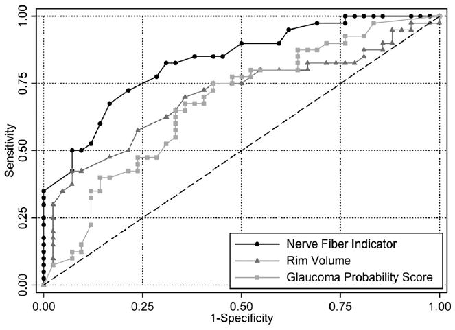
Main outcome measures: Areas under the receiver operating characteristic (ROC) curves were used to evaluate the diagnostic accuracies of GDx VCC and the HRT.
Results: Forty eyes with progressive glaucomatous optic nerve change were included in the glaucoma group, and 42 eyes without any evidence of progressive damage to the optic nerve followed untreated for an average time of 8.97+/-3.08 years were included in the normal group. The area under the ROC curve for the best parameter from GDx VCC (nerve fiber indicator [NFI]) was significantly larger than that of the best parameter from the HRT (rim volume) (0.83 vs. 0.70; P = 0.044). The NFI parameter also had a larger ROC curve area than that of the contour line-independent parameter glaucoma probability score (0.83 vs. 0.68; P = 0.023). Assuming borderline results as normal, the Moorfields regression analysis classification had a sensitivity of 48% for specificity of 69%. For a similar specificity (70%), the parameter NFI had a significantly larger sensitivity (83%) (P = 0.003).
Conclusions: Retinal nerve fiber layer imaging with GDx VCC had a superior performance versus topographic optic disc assessment with the HRT for detecting early damage in patients suspected of having glaucoma. For glaucoma diagnosis, these results suggest that GDx VCC may offer advantage over the HRT when these tests are combined with clinical examination of the optic nerve.
Figures
Similar articles
-
Detection of psychophysical and structural injury in eyes with glaucomatous optic neuropathy and normal standard automated perimetry.Arch Ophthalmol. 2006 Feb;124(2):169-76. doi: 10.1001/archopht.124.2.169. Arch Ophthalmol. 2006. PMID: 16476885
-
Optic disk and nerve fiber layer imaging to detect glaucoma.Am J Ophthalmol. 2007 Nov;144(5):724-32. doi: 10.1016/j.ajo.2007.07.010. Epub 2007 Sep 14. Am J Ophthalmol. 2007. PMID: 17868631 Free PMC article.
-
Optic disc imaging in perimetrically normal eyes of glaucoma patients with unilateral field loss.Trans Am Ophthalmol Soc. 2006;104:202-11. Trans Am Ophthalmol Soc. 2006. PMID: 17471341 Free PMC article.
-
Retinal nerve fiber layer analysis in the diagnosis of glaucoma.Curr Opin Ophthalmol. 2006 Apr;17(2):120-31. doi: 10.1097/01.icu.0000193079.55240.18. Curr Opin Ophthalmol. 2006. PMID: 16552246 Review.
-
Role of optic nerve imaging in glaucoma clinical practice and clinical trials.Am J Ophthalmol. 2008 Apr;145(4):598-603. doi: 10.1016/j.ajo.2007.12.018. Epub 2008 Mar 4. Am J Ophthalmol. 2008. PMID: 18295183 Free PMC article. Review.
Cited by
-
Detection of progressive retinal nerve fiber layer loss in glaucoma using scanning laser polarimetry with variable corneal compensation.Invest Ophthalmol Vis Sci. 2009 Apr;50(4):1675-81. doi: 10.1167/iovs.08-2712. Epub 2008 Nov 21. Invest Ophthalmol Vis Sci. 2009. PMID: 19029038 Free PMC article.
-
A combined index of structure and function for staging glaucomatous damage.Arch Ophthalmol. 2012 Sep;130(9):1107-16. doi: 10.1001/archophthalmol.2012.827. Arch Ophthalmol. 2012. PMID: 23130365 Free PMC article.
-
Advancement in Understanding Glaucoma: A Comprehensive Review.Cureus. 2023 Sep 30;15(9):e46254. doi: 10.7759/cureus.46254. eCollection 2023 Sep. Cureus. 2023. PMID: 37908941 Free PMC article. Review.
-
A simplified combined index of structure and function for detecting and staging glaucomatous damage.Sci Rep. 2021 Feb 4;11(1):3172. doi: 10.1038/s41598-021-82756-6. Sci Rep. 2021. PMID: 33542367 Free PMC article.
-
Glaucoma Diagnosis and Monitoring Using Advanced Imaging Technologies.US Ophthalmic Rev. 2013;6(1):15-25. US Ophthalmic Rev. 2013. PMID: 24470807 Free PMC article.
References
-
- Weinreb RN, Shakiba S, Zangwill L. Scanning laser polarimetry to measure the nerve fiber layer of normal and glaucomatous eyes. Am J Ophthalmol. 1995;119:627–36. - PubMed
-
- Medeiros FA, Zangwill LM, Bowd C, et al. Comparison of scanning laser polarimetry using variable corneal compensation and retinal nerve fiber layer photography for detection of glaucoma. Arch Ophthalmol. 2004;122:698–704. - PubMed
-
- Weinreb RN. Assessment of optic disc topography for diagnosing and monitoring glaucoma. Arch Ophthalmol. 1998;116:1229–31. - PubMed
-
- Medeiros FA, Zangwill LM, Bowd C, Weinreb RN. Comparison of the GDx VCC scanning laser polarimeter, HRT II confocal scanning laser ophthalmoscope, and Stratus OCT optical coherence tomograph for the detection of glaucoma. Arch Ophthalmol. 2004;122:827–37. - PubMed
-
- Medeiros FA, Zangwill LM, Bowd C, et al. Influence of disease severity and optic disc size on the diagnostic performance of imaging instruments in glaucoma. Invest Ophthalmol Vis Sci. 2006;47:1008–15. - PubMed
Publication types
MeSH terms
Grants and funding
LinkOut - more resources
Full Text Sources
Medical
