Review
. 2008 Apr;84(4):363-404.
doi: 10.1016/j.pneurobio.2007.10.010. Epub 2007 Dec 8.

Progress in neuroprotective strategies for preventing epilepsy

Affiliations
Review

Progress in neuroprotective strategies for preventing epilepsy

Munjal M Acharya et al. Prog Neurobiol. 2008 Apr.

Abstract

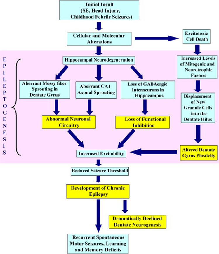
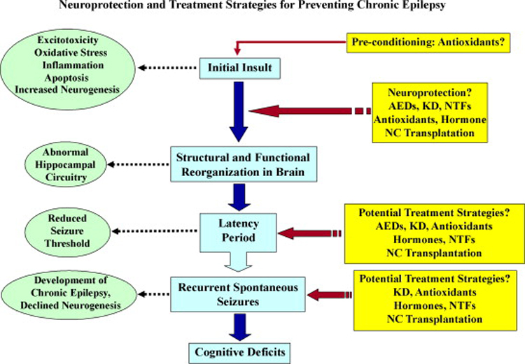
Neuroprotection is increasingly considered as a promising therapy for preventing and treating temporal lobe epilepsy (TLE). The development of chronic TLE, also termed as epileptogenesis, is a dynamic process. An initial precipitating injury (IPI) such as the status epilepticus (SE) leads to neurodegeneration, abnormal reorganization of the brain circuitry and a significant loss of functional inhibition. All of these changes likely contribute to the development of chronic epilepsy, characterized by spontaneous recurrent motor seizures (SRMS) and learning and memory deficits. The purpose of this review is to discuss the current state of knowledge pertaining to neuroprotection in epileptic conditions, and to highlight the efficacy of distinct neuroprotective strategies for preventing or treating chronic TLE. Although the administration of certain conventional and new generation anti-epileptic drugs is effective for primary neuroprotection such as reduced neurodegeneration after acute seizures or the SE, their competence for preventing the development of chronic epilepsy after an IPI is either unknown or not promising. On the other hand, alternative strategies such as the ketogenic diet therapy, administration of distinct neurotrophic factors, hormones or antioxidants seem useful for preventing and treating chronic TLE. However, long-term studies on the efficacy of these approaches introduced at different time-points after the SE or an IPI are lacking. Additionally, grafting of fetal hippocampal cells at early time-points after an IPI holds considerable promise for preventing TLE, though issues regarding availability of donor cells, ethical concerns, timing of grafting after SE, and durability of graft-mediated seizure suppression need to be resolved for further advances with this approach. Overall, from the studies performed so far, there is consensus that neuroprotective strategies need to be employed as quickly as possible after the onset of the SE or an IPI for considerable beneficial effects. Nevertheless, ideal strategies that are capable of facilitating repair and functional recovery of the brain after an IPI and preventing the evolution of IPI into chronic epilepsy are still hard to pin down.


Figures

Figure 1
Figure 1
The various aspects of epileptogenesis after the initial brain insult and the evolution of the initial precipitating injury into chronic epilepsy and learning and memory deficits. An initial insult in the form of head injury or the status epilepticus (SE) typically leads to a number of cellular and molecular changes in the hippocampus. A transient surge in the proliferation of neural stem/progenitor cells also occurs in the dentate gyrus immediately after the SE resulting in abnormal neurogenesis. A multitude of alterations in the milieu of the dentate gyrus and the hippocampal CA1 and CA3 subfields lead to abnormal synaptic reorganization, the loss of functional inhibition by the GABAergic system and altered dentate gyrus plasticity, all of which augment the process of epileptogenesis. Collectively, these changes contribute to the occurrence of spontaneous recurrent motor seizures (SRMS) and learning and memory deficits during the chronic phase of epilepsy.
Figure 2
Figure 2
Neurodegeneration after the status epilepticus in hippocampal and extrahippocampal regions. The degenerating neurons were visualized at 24 hr after the status epilepticus through Fluoro-Jade B (A1-B5), silver (C1-D4), and TUNEL staining (E1-E3) in different regions of the hippocampus, amygdala, and the entorhinal cortex. Scale bars, A1-A3 and B1-B3 = 100µm; A4, A5, B4, B5 = 50µm; C1-C3 and D1-D4 = 20m; E = 200 µm. (Figure reproduced from: Rao et al., 2006; J Neurosci Res. 83(6):1088–1105).
Figure 3
Figure 3
Aberrant mossy fiber sprouting after the status epilepticus in the dentate gyrus. The extent of the aberrant mossy fiber sprouting is illustrated for rats with moderate hippocampal injury (B1, B2 and E1, E2) and rats with severe hippocampal injury (C1,C2 and F1,F2), in comparison to age-matched intact rats (A1,2 and D1,D2) by Timm's histochemical staining (A1-C2) and neuropeptide Y (NPY) immunostaining (D1-F2). Note that, in comparison to rats exhibiting moderate hippocampal injury (B1, B2 and E1, E2), rats with severe hippocampal injury (C1, C2 and F1, F2) exhibit much robust aberrant sprouting of mossy fibers into the dentate supragranular layer (DSGL). DH, dentate hilus; GCL, granule cell layer. Scale bars, A, B1, C1 = 500 µm; A2, B2, C2 = 100 µm; D1, E1 and F1 = 500µm; D2, E2, F2 = 200 µm. (Figures reproduced from: Rao et al., 2006; J Neurosci Res. 83(6):1088–1105).
Figure 4
Figure 4
Top left panel: Morphology of the biotinylated dextran amine (BDA)-positive entorhinal axons in the CA3-lesioned hippocampus at 3 months after an intracerebroventricular administration of the kainic acid. A1, A2: Entorhinal axon of the alvear pathway traversing the CA1 stratum radiatum in an intact hippocampus showing wavy axons exhibiting a large number of en passant bouton (arrowheads). B1-B4: Region of the CA1 subfield from a CA3-lesioned hippocampus exhibiting a large number of horizontally oriented axons (arrows) filled with boutons (arrowheads), and branches (arrow in B3) and growth cone-like expansions at their termination in the outer thirds of the CA1 stratum radiatum. GCL, granule cell layer; IML, inner molecular layer; MML, middle molecular layer; OML, outer molecular layer; SLM, stratum lacunosum moleculare; SO, stratum oriens; SP, stratum pyramidale; SR, stratum radiatum. Scale bars, A1-A3, B2-B4 and C2 = 10 µm; B1 = 50 µm. (Figure reproduced from: Shetty, AK; 2002; Hippocampus; 12(4):534–542). Top right panel: Loss of GABA-ergic interneurons following kainic acid (KA) induced hippocampal injury. Note that KA induced injury reduces the density of GABA-ergic interneurons in the dentate gyrus, and CA1 and CA3 subfields (B1-B4), in comparison to the density typically observed in these regions of the naïve hippocampus (A1-A4). GCL, granule cell layer; DH, dentate hilus; SLM, stratum lacunosum moleculare; SO, stratum oriens; SP, stratum pyramidale; SR, stratum radiatum. (Figure reproduced from Shetty AK, Turner DA, J. Neurosci. 2006). Bottom panel: Hippocampal cytoarchitecture and distribution of newly formed doublecortin (DCX) immunopositive neurons in the dentate gyrus following intraperitoneal kainic acid (IPKA) injections. The photographs A1 and A2 show Nissl-stained sections from the septal and temporal regions of the hippocampus showing milder (A1) and severe (A2) neurodegeneration (Asterisks). The photographs B1, B2, B3 show the distribution of dramatically increased DCX immunopositive new neurons in the dentate gyrus at 16 days after IP KA injections. The photographs C1, C2, C3 show severely declined dentate neurogenesis (as revealed by only a few DCX immunopositive new neurons) at 5 months after KA injections. Scale bars, A1, A2 = 500 µm; B1, C1 = 200µm; B2, B3, C2 and C3 = 50µm. (Reproduced from: Hattiangady et al., 2004; Neurobiol Dis. 17(3):473–490).
Figure 5
Figure 5
A schematic representation of the metabolism of glucose, ketone bodies and amino acids (excitatory neurotransmitters) in the brain. Approximately 90% of dietary calories derived from fats induce ketosis through fatty acid metabolism in the liver. The major ketone bodies produced by this route comprise 3-hydroxy-butyrate (3-OH-butyrate) and acetoacetate, which serve as fuel for high-energy demand of the brain in epileptic conditions. The ketone body acetoacetate is sequestered to acetoacetyl-CoA by succinyl-CoA transferase (SCOT) in the brain. In parallel, the pyruvate (through glycolysis) generates acetyl-CoA by pyruvate dehydrogenase complex (PDH) and a fraction of lactate through lactate dehydrogenase (LDH). The acetoacetyl-CoA routed via ketone bodies metabolism also generates excess pool of acetyl-CoA in the mitochondrial acetoacetyl-CoA thiolase (AACOT) reaction and enters the tricarboxylic acid (TCA) cycle. Hence, oxaloacetate (OAA) pool diminishes because of increased availability of substrates for key TCA cycle enzyme citrate synthetase (CS). As a result, less OAA is available for transamination reaction of aspartate aminotransferase (AAT) to produce aspartate, which in turn leads to increased glutamate pool for glutamic acid decarboxylase (GAD) and favors GABA synthesis. Alternatively, glutamine (Gln) production would likely reduce the glutamate load in neurons.
Figure 6
Figure 6
The potential role of ketogenic diet on mitochondrial dependent apoptotic signaling cascades. The ketogenic diet therapy is associated with increased activity of anti-apoptotic proteins like Akt and molecular chaperon 14-3-3 and reduced activity of pro-apoptotic proteins Bad, Bax and caspase-3. Akt phosphorylates Bad that forms a complex with 14-3-3. This prevents further activation of Bax that is involved in the mitochondrial permeability transition pore (MPT) formation and subsequent release of cytochrome c (cyt c) into the cytosol. Overall, the ketogenic diet helps to prevent the caspase-dependent apoptotic cell death.
Figure 7
Figure 7
Concentration of Brain derived neurotrophic factor (BDNF) in hippocampi ipsi- and contralateral to unilateral kainic acid (KA) administration at different time-points after the KA administration. Note that the BDNF is significantly up-regulated at 4 days post-KA administration, reaches the level observed in the intact hippocampus at 45 days post-KA, and decreases below the control levels at 120 days post-KA. The hippocampus contralateral to KA administration retains baseline BDNF until 45 days post lesion but exhibits considerable decline at 120 day post-KA. (Figure reproduced from:Shetty et al., 2003; J Neurochem; 87:147–159).
Figure 8
Figure 8
Top panel: Survival and morphology of fetal CA3 cell grafts placed close to the lesioned CA3 region of the adult hippocampus (marked by asterisks) at 45 days post-lesion and analyzed at 1 year postgrafting. A1 and A2 show a Nissl-stained section of the rat hippocampus containing fetal CA3 transplant (outlined by asterisks) located just below the degenerated CA3 cell layer (indicated by interrupted lines). B1, B2 show NeuN positive neurons within the graft in a neighboring section. DH, dentate hilus. Scale bar: A1, B1 = 400µm; A2, B2 = 50µm. (Reproduced from: Zaman, V and Shetty, AK; 2001; Neurobiol Dis.; 8:942–95). Bottom panel: Effects of CA3 or CA1 cell grafts on aberrant mossy fiber sprouting analyzed at one year post- grafting. Note dense mossy fiber sprouting in the dentate supragranular layer in the lesion only group (A1, D2) and a marked reduction in mossy fiber sprouting (B1, D3) following grafting of fetal CA3 cells but not with CA1 cells (C1, D4). B2 shows dense innervation of fetal CA3 cells grafted into the degenerated host CA3 region by host mossy fibers. C2 shows lack of host mossy fiber growth into the fetal CA1 graft. DH, dentate hilus. Scale bar: Scale bar: A1, B1, C1=500µm; B2, C2, D1–4 = 100µm.
Figure 9
Figure 9
Potential neuroprotection and treatment strategies for preventing the chronic epilepsy development after an initial precipitating injury. An IPI leads to abnormal structural and functional reorganization of the brain circuitry, which gradually develops into chronic epilepsy characterized by spontaneous recurrent motor seizures (SRMS). A number of molecular as well as organizational level alterations occur during this gradual process. The neuroprotective interventions such as administration of the anti-epileptic drugs (AEDs), the ketogenic diet therapy (KD), the neurotrophic factors such as GDNF and NT-3, the antioxidants such as resveratrol and Curcumin and transplants of cells containing fetal hippocampal cells or neural stem cells may be beneficial immediately after an IPI for preventing subsequent epileptogenesis. The potential treatment strategies either during or after latency periods may also help in controlling the SRMS and cognitive deficits. Additionally, pre-conditioning with natural anti-oxidants may be effective in reducing IPI or seizure induced brain damage and the consequent epileptogenesis.

References

    1. Acharya MM, Katyare SS. Structural and functional alterations in mitochondrial membrane in picrotoxin-induced epileptic rat brain. Exp Neurol. 2005;192:79–88. - PubMed
    1. Acuna-Castroviejo D, Escames G, Macias M, Munoz Hoyos A, Molina Carballo A, Arauzo M, Montes R. Cell protective role of melatonin in the brain. J Pineal Res. 1995;19:57–63. - PubMed
    1. Adams B, Sazgar M, Osehobo P, Van der Zee CE, Diamond J, Fahnestock M, Racine RJ. Nerve growth factor accelerates seizure development, enhances mossy fiber sprouting, and attenuates seizure-induced decreases in neuronal density in the kindling model of epilepsy. J Neurosci. 1997;17:5288–5296. - PMC - PubMed
    1. Airaksinen MS, Saarma M. The GDNF family: signalling, biological functions and therapeutic value. Nature Rev. 2002;3:383–394. - PubMed
    1. Ajmone-Cat MA, Iosif RE, Ekdahl CT, Kokaia Z, Minghetti L, Lindvall O. Prostaglandin E2 and BDNF levels in rat hippocampus are negatively correlated with status epilepticus severity: no impact on survival of seizure-generated neurons. Neurobiol Dis. 2006;23:23–35. - PubMed

Publication types

MeSH terms