

Mucosal transmission of R5 and X4 tropic HIV-1 via vaginal and rectal routes in humanized Rag2-/- gammac -/- (RAG-hu) mice
- PMID: 18207484
- PMCID: PMC3092740
- DOI: 10.1016/j.virol.2007.11.020
Mucosal transmission of R5 and X4 tropic HIV-1 via vaginal and rectal routes in humanized Rag2-/- gammac -/- (RAG-hu) mice
Abstract
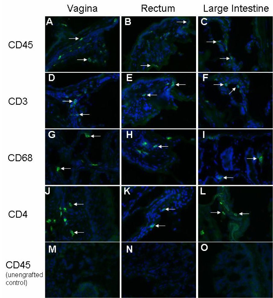
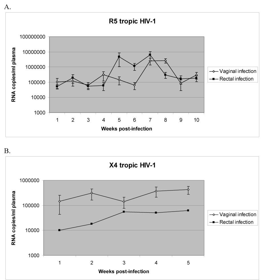
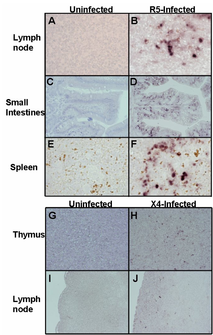
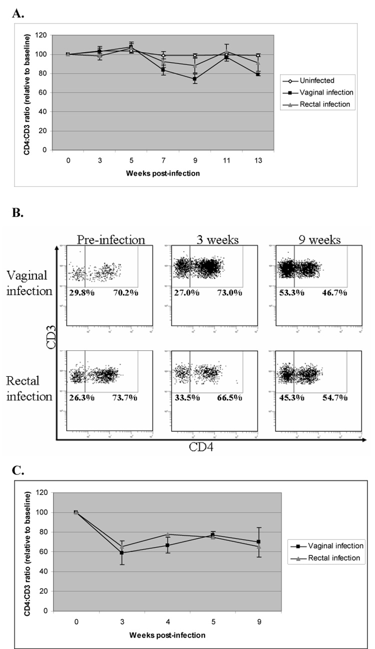
Studies on HIV-1 mucosal transmission to evaluate early events in pathogenesis and the development of effective preventive/prophylactic methods have thus far been hampered by the lack of a suitable animal model susceptible to HIV-1 infection by either vaginal and/or rectal routes. In this regard, while primate-SIV/SHIV and cat-FIV models provided useful surrogate platforms to derive comparative data, these viruses are distinct and different from that of HIV-1. Therefore an optimal model that permits direct study of HIV-1 transmission via mucosal routes is highly desirable. The new generation of humanized NOD/SCID BLT, NOD/SCIDgammac(-/-), and Rag2(-/-)gammac(-/-) mouse models show great promise to achieve this goal. Here, we show that humanized Rag2(-/-)gammac(-/-) mice (RAG-hu) engrafted with CD34 hematopoietic progenitor cells harbor HIV-1-susceptible human cells in the rectal and vaginal mucosa and are susceptible to HIV-1 infection when exposed to cell-free HIV-1 either via vagina or rectum. Infection could be established without any prior hormonal conditioning or mucosal abrasion. Both R5 and X4 tropic viruses were capable of mucosal infection resulting in viremia and associated helper T cell depletion. There was systemic spread of the virus with infected cells detected in different organs including the intestinal mucosa. R5 virus was highly efficient in mucosal transmission by both routes whereas X4 virus was relatively less efficient in causing infection. HIV-1 infection of RAG-hu mice by vaginal and rectal routes as shown here represents the first in vivo model of HIV-1 transmission across intact mucosal barriers and as such may prove very useful for studying early events in HIV-1 pathogenesis in vivo, as well as the testing of microbicides, anti-HIV vaccines/therapeutics, and other novel strategies to prevent HIV-1 transmission.
Figures
References
-
- Anderson J, Akkina R. Complete knockdown of CCR5 by lentiviral vector-expressed siRNAs and protection of transgenic macrophages against HIV-1 infection. Gene Ther. 2007;14:1287–1297. - PubMed
-
- Baenziger S, Tussiwand R, Schlaepfer E, Mazzucchelli L, Heikenwalder M, Kurrer MO, Behnke S, Frey J, Oxenius A, Joller H, Aguzzi A, Manz MG, Speck RF. Disseminated and sustained HIV infection in CD34+ cord blood cell-transplanted Rag2−/−{gamma}c−/− mice. Proc Natl Acad Sci USA. 2006;103:15951–15956. - PMC - PubMed
-
- Berger EA, Murphy PM, Farber JM. Chemokine receptors as HIV-1 coreceptors: roles in viral entry, tropism, and disease. Ann Rev Imm. 1999;17:657–700. - PubMed
Publication types
MeSH terms
Substances
Grants and funding
LinkOut - more resources
Full Text Sources
Other Literature Sources
Medical
Miscellaneous
