

Augmented serum level of major histocompatibility complex class I-related chain A (MICA) protein and reduced NKG2D expression on NK and T cells in patients with cervical cancer and precursor lesions
- PMID: 18208618
- PMCID: PMC2270854
- DOI: 10.1186/1471-2407-8-16
Augmented serum level of major histocompatibility complex class I-related chain A (MICA) protein and reduced NKG2D expression on NK and T cells in patients with cervical cancer and precursor lesions
Abstract
Background: Cervical cancer is the second most common cancer in women worldwide. NK and cytotoxic T cells play an important role in the elimination of virus-infected and tumor cells through NKG2D activating receptors, which can promote the lysis of target cells by binding to the major histocompatibility complex class I-related chain A (MICA) proteins. Increased serum levels of MICA have been found in patients with epithelial tumors. The aim of this study was to compare the levels of soluble MICA (sMICA) and NKG2D-expressing NK and T cells in blood samples from patients with cervical cancer or precursor lesions with those from healthy donors.
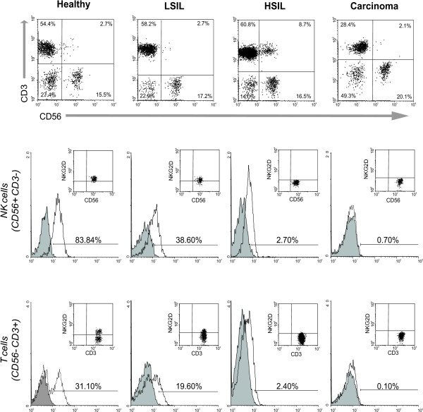
Methods: Peripheral blood with or without heparin was collected to obtain mononuclear cells or sera, respectively. Serum sMICA levels were measured by ELISA and NKG2D-expressing immune cells were analyzed by flow cytometry. Also, a correlation analysis was performed to associate sMICA levels with either NKG2D expression or with the stage of the lesion.
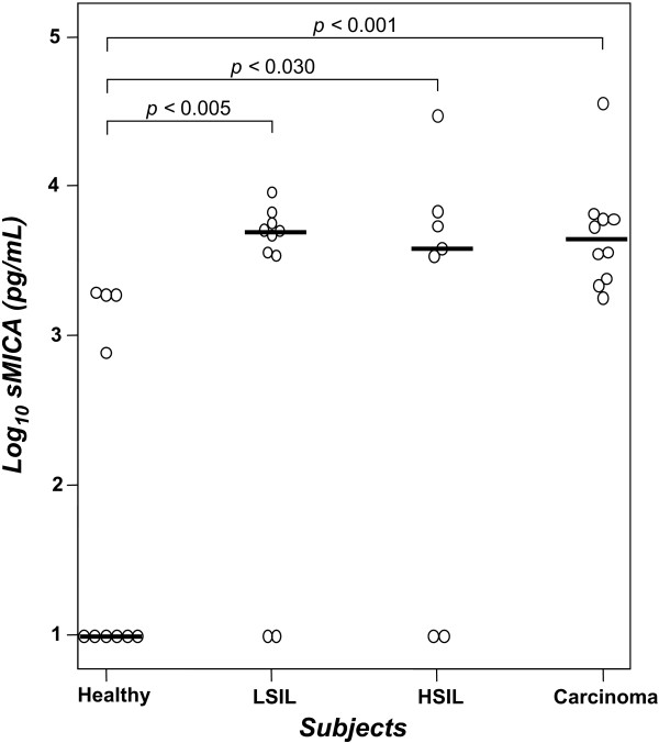
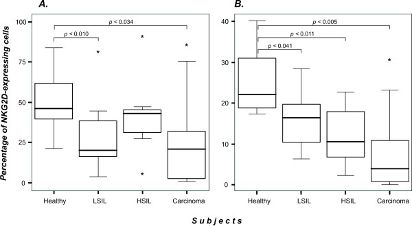
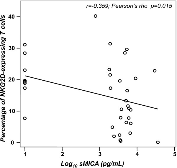
Results: Significant amounts of sMICA were detected in sera from nearly all patients. We found a decrease in the number of NKG2D-expressing NK and T cells in both cervical cancer and lesion groups when compared to healthy donors. Pearson analysis showed a negative correlation between sMICA and NKG2D-expressing T cells; however, we did not find a significant correlation when the analysis was applied to sMICA and NKG2D expression on NK cells.
Conclusion: Our results show for the first time that high sMICA levels are found in sera from patients with both cervical cancer and precursor lesions when compared with healthy donors. We also observed a diminution in the number of NKG2D-expressing NK and T cells in the patient samples; however, a significant negative correlation between sMICA and NKG2D expression was only seen in T cells.
Figures
References
Publication types
MeSH terms
Substances
LinkOut - more resources
Full Text Sources
Other Literature Sources
Medical
