. 2008 Feb 1;180(3):1758-68.
doi: 10.4049/jimmunol.180.3.1758.

Healthy human subjects have CD4+ T cells directed against H5N1 influenza virus

Affiliations

Healthy human subjects have CD4+ T cells directed against H5N1 influenza virus

Michelle Roti et al. J Immunol. .

Abstract

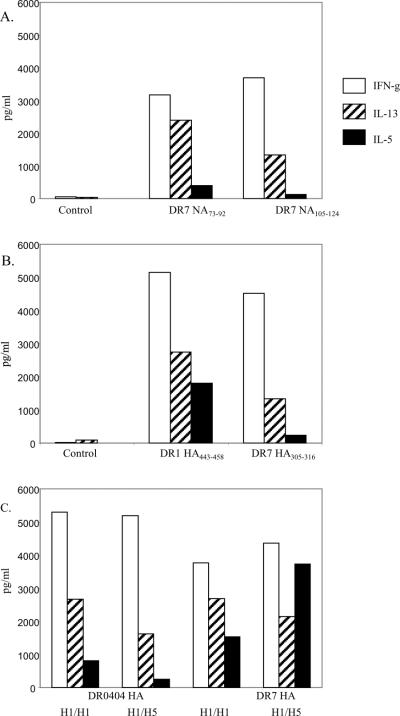
It is commonly perceived that the human immune system is naive to the newly emerged H5N1 virus. In contrast, most adults have been exposed to influenza A H1N1 and H3N2 viruses through vaccination or infection. Adults born before 1968 have likely been exposed to H2N2 viruses. We hypothesized that CD4(+) T cells generated in response to H1N1, H3N2, and H2N2 influenza A viruses also recognize H5N1 epitopes. Tetramer-guided epitope mapping and Ag-specific class II tetramers were used to identify H5N1-specific T cell epitopes and detect H5N1-specific T cell responses. Fifteen of 15 healthy subjects tested had robust CD4(+) T cell responses against matrix protein, nucleoprotein, and neuraminidase of the influenza A/Viet Nam/1203/2004 (H5N1) virus. These results are not surprising, because the matrix protein and nucleoprotein of influenza A viruses are conserved while the neuraminidase of the H5N1 virus is of the same subtype as that of the circulating H1N1 influenza strain. However, H5N1 hemagglutinin-reactive CD4(+) T cells were also detected in 14 of 14 subjects examined despite the fact that hemagglutinin is less conserved. Most were cross-reactive to H1, H2, or H3 hemagglutinin epitopes. H5N1-reactive T cells were also detected ex vivo, exhibited a memory phenotype, and were capable of secreting IFN-gamma, TNF-alpha, IL-5, and IL-13. These data demonstrate the presence of H5N1 cross-reactive T cells in healthy Caucasian subjects, implying that exposure to influenza A H1N1, H3N2, or H2N2 viruses through either vaccination or infection may provide partial immunity to the H5N1 virus.


Figures

Figure 1
Figure 1
Identification of DR0404 restricted M1 epitopes. CD4+ T cells from a DR0404 subject were stimulated with 6 peptide pools derived from the M1 protein of A/Viet Nam/1203/2004. A. Cells were stained with the corresponding DR0404 pooled M1 peptide tetramers at day 14. Pools 2, 3, 4 and 6 gave positive staining. B. Cells stimulated with peptide pools that gave positive staining were stained with DR0404 individual peptide tetramers at day 17. The percentage of tetramer positive cells is indicated. Background staining in these experiments was 0.3% or lower. Peptides p9, p10, p13, p16, p26 and p27, which correspond to M165–84, M173–92, M197–116, M1121–140, M1201–220 and M1209–228 respectively, gave staining significantly above background and were identified as H5 M1 epitopes.
Figure 2
Figure 2
Ex vivo staining of H5 M1 and H5 NP reactive T cells. PBMC from DR0101, DR0404 DR0701 and DR1101 subjects were stained with a panel of HLA- matched DR0101/M1, DR0101/NP, DR0404/M1, DR0404/NP, DR0701/M1, DR0701/NP, DR1101/M1, DR1101/NP tetramers or the appropriate control tetramers. The panel of tetramers being used and the ex vivo staining protocol were as stated in the text. The DR0101 control tetramers was DR0101/GAD65555–567, the DR0404 control tetramers was DR0404/GAD65555–567, and both the DR0701 and DR1101 control tetramers were empty tetramers. The frequency of antigen specific T cell per million CD4+ T cells is as indicated.
Figure 3
Figure 3
Ex vivo staining of NA reactive T cells. PBMC from DR0101, DR0404, DR0701 and DR1101 subjects were stained with a panel of HLA- matched NA tetramers. For the DR0101 subject, the tetramer was DR0101/NA105–124. For the DR0404 subject, the tetramers were DR0404/NA345–364 and DR0404/NA409–428. For the DR0701 subject, the tetramers were DR0701/NA73–92 and DR0701/NA105–124. For the DR1101 subject, the tetramers were DR1101/NA225–244 and DR1101/NA393–412. The frequency of antigen specific T cell per million CD4+ T cells is as indicated.
Figure 4
Figure 4
Identification of DR0404 restricted HA epitopes. CD4+ T cells from a DR0404 subject were stimulated with 14 peptide pools derived from the HA protein of A/Viet Nam/1203/2004. A. Cells were stained with the corresponding DR0404 pooled HA peptide tetramers at day 14. Pools 5, 10, 11 and 12 were identified as positive pools. B. Cells stimulated with peptide pools that gave positive staining were stained with DR0404 individual peptide tetramers at day 17. The percentage of tetramer positive cells is indicated. Background staining in these experiments was 0.3% or lower. Peptides p22, p50, p51 and p56, which correspond to HA169–188, HA393–412, HA401–420 and HA441–460 respectively, were identified as T cell epitopes.
Figure 5
Figure 5
H5N1 HA CD4+ T cells are memory CD4+ T cells. CD4+ T cells were sorted into CD45RA+ and CD45RA− fractions using a FACSVantage. The CD4+CD45RA− fraction was stimulated with H5 HA peptides. A. Cells from the CD45RA− fraction of a DR0701 subject were stimulated with HA253–265 (middle panel) or HA305–316 peptide (right panel). At day 14, these cells were stained with the corresponding DR0701 tetramers. The negative control was cells which have been stimulated with a M133–52 peptide and stained with DR0701/ HA253–265 tetramers. B. Cells from the CD45RA− fraction of a DR1101 subject were stimulated with HA267–279 (middle panel) or HA319–331 peptide (right panel). At day 14, these cells were stained with the corresponding DR1101 tetramers. The negative control was cells that have been stimulated with M197–116, and stained with DR1101/ HA267–279 tetramers.
Figure 6
Figure 6
Ex vivo staining of H5 HA reactive T cells. A. PBMC from DR0101, DR0404, DR0701, DR1101 and DR1501 subjects were stained with a panel of HLA-matched DR0101/HA, DR0404/HA, DR0701/HA, DR1101/HA and DR1501/HA tetramers. The tetramers used were as followed, DR0101/ HA91–104, DR0101/ HA157–169 and DR0101/ HA443–458 for DR0101 subjects, DR0404/ HA174–188, DR0404/ HA400–412 and DR0404/ HA443–458 for DR0404 subjects, DR0701/ HA49–68, DR0701/ HA253–265 and DR0701/ HA305–316 for DR0701 subjects, D1101/ HA267–279, DR1101/ HA319–331 and DR1101/ HA408–420 for DR1101 subjects, and DR1501/ HA433–452 for DR1501 subjects. B. As a negative control for the tetramer staining, PBMC from a DR0404 subject was stained with the same DR1501/HA tetramers used in A, and PBMC from a DR1501 subject was stained with the set of DR0404/HA tetramers described as in A. The frequency of antigen specific T cell per million CD4+ T cells is as indicated. No significant staining was observed in the DR1101 subject in A. and the two negative controls in B.
Figure 7
Figure 7
Cross-reactivity of H5 HA253–265 and HA305–316 reactive T cells. A. CD4+ T cells from a DR0701 subject were stimulated with H5 HA253–265, or with peptides in the corresponding region of H1N1, H2N2 or H3N2 HA (sequences are as indicated in Table IV). Fourteen days post-stimulation, cells were stained with DR0701/ H5 HA253–265 tetramers. B. CD4+ T cells from a DR0701 subject were stimulated with H5 HA305–316, or with peptides in the corresponding region of H1N1, H2N2 or H3N2 HA (sequences are as indicated in Table IV). Fourteen days post-stimulation, cells were stained with DR0701/ H5 HA305–316 tetramers. C. CD4+ T cells from a DR0404 subject were stimulated with H5 HA408–420, or with peptides in the corresponding regions of H1N1 or H3N2 HA (see Table IV). Fourteen days post-stimulation, cells were stained with DR0404/ H5 HA408–420 tetramers. The aa sequences of H5 HA408–420 and the corresponding region of H2 HA are exactly identical.
Figure 8
Figure 8
Ex vivo cytokine analysis of H5N1 reactive T cells. DR0404 restricted H5N1 M1, NP, NA, and HA T cells were analysis for secretion of IFN-γ and TNF-α after an overnight culture of the PBMC from a DR0404 subject with a panel of M1, NP, NA or HA specific tetramers. The DR0404 M1, NP, NA and HA tetramers used were as previously described. The DR0404/GAD65555–567 tetramers were used as a negative control. The frequencies of IFN-γ and TNF-α secreting cells per million CD4+ T cells are as indicated.
Figure 9
Figure 9
Cytokine profiles of NA and HA reactive T cells. A. Cytokine profiles of NA reactive T cells. PBMC from a DR0701 subject were stimulated with the NA73–92 (middle panel) or the NA105–124 (right panel) peptide. On day 14, cells were transferred to a well that were precoated with the corresponding tetramers, and supernatants were harvested after an overnight culture. Cytokines were assayed by the Meso Scale multiplex kit. Cells had been stimulated with the NA105–124 peptide were also reactivated with an irrelevant tetramer as a negative control (panel on the left). B. Cytokine profiles of HA reactive T cells. PBMC from a DR0101 subject (middle panel) and a DR0701 subject (right panel) were stimulated with the H5 HA443–458 peptide and the H5 HA305–316 peptide respectively. Cytokines were assayed by the Meso Scale multiplex kit as described above. Cells from the DR0101 subjects were also restimulated with an irrelevant tetramer as a negative control (left panel). C. Cross-reactivity of H1 and H5 HA reactive T cells. PBMC from a DR0404 subject were stimulated with the H1 HA peptide SVIEKMNTQFTAV (Table IV). Cells were reactivated with either the H1 HA peptide or the H5 HA400–412 peptide and then assayed for cytokines (first set and second set of data respectively). PBMC from a DR0701 subject were stimulated with the H1 HA peptide DTIIFEANGNLIA (Table IV). Cells were reactivated with either the H1 HA peptide or the H5 HA253–265 peptide and then assayed for cytokines (third and fourth set of data respectively).

References

    1. Olsen B, Munster VJ, Wallensten A, Waldenstrom J, Osterhaus AD, Fouchier RA. Global patterns of influenza a virus in wild birds. Science. 2006;312:384–388. - PubMed
    1. Webster RG, Peiris M, Chen H, Guan Y. H5N1 outbreaks and enzootic influenza. Emerg. Infect. Dis. 2006;12:3–8. - PMC - PubMed
    1. Woo PC, Lau SK, Yuen KY. Infectious diseases emerging from Chinese wet-markets: zoonotic origins of severe respiratory viral infections. Curr. Opin. Infect. Dis. 2006;19:401–407. - PMC - PubMed
    1. Beigel JH, Farrar J, Han AM, Hayden FG, Hyer R, de Jong MD, Lochindarat S, Nguyen TK, Nguyen TH, Tran TH, Nicoll A, Touch S, Yuen KY. Avian influenza A (H5N1) infection in humans. N. Engl. J. Med. 2005;353:1374–1385. - PubMed
    1. Wong SS, Yuen KY. Avian influenza virus infections in humans. Chest. 2006;129:156–168. - PMC - PubMed

Publication types