. 2008 Feb 1;180(3):1843-53.
doi: 10.4049/jimmunol.180.3.1843.

Cyclooxygenase-2 deficiency enhances Th2 immune responses and impairs neutrophil recruitment in hepatic ischemia/reperfusion injury

Affiliations

Cyclooxygenase-2 deficiency enhances Th2 immune responses and impairs neutrophil recruitment in hepatic ischemia/reperfusion injury

Takashi Hamada et al. J Immunol. .

Abstract

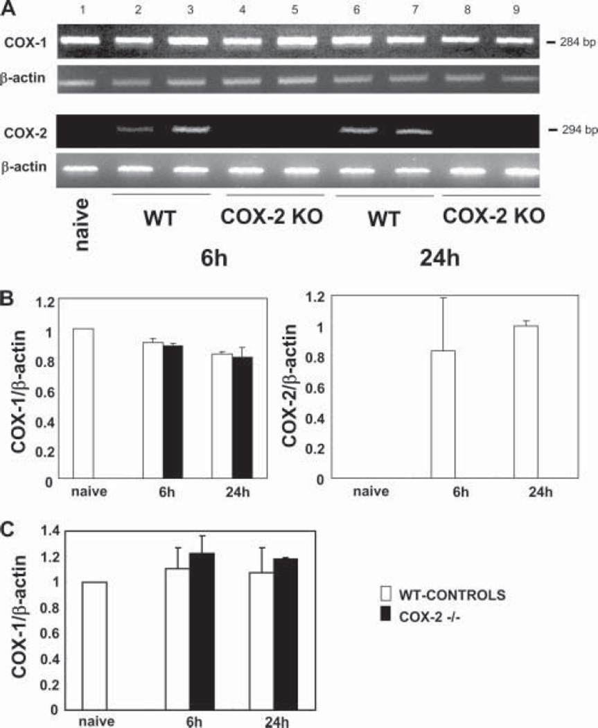
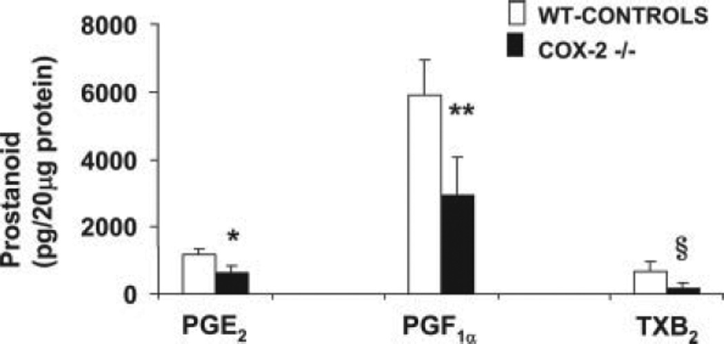
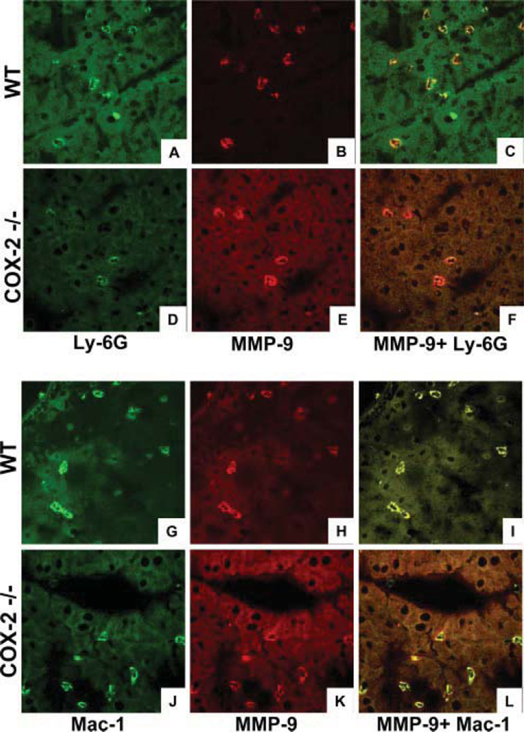
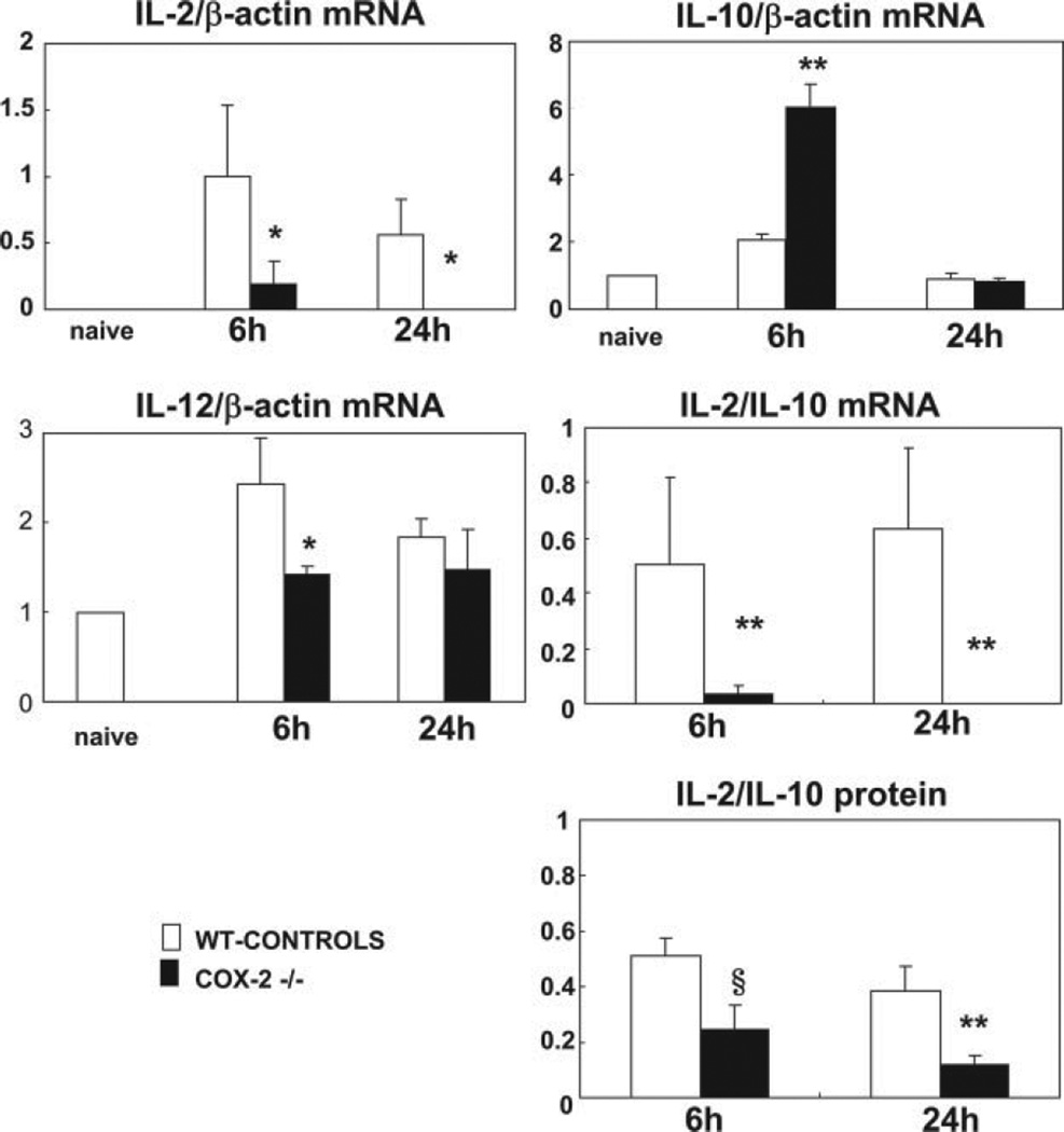
Cyclooxygenase-2 (COX-2) is a prostanoid-synthesizing enzyme that is critically implicated in a variety of pathophysiological processes. Using a COX-2-deficient mouse model, we present data that suggest that COX-2 has an active role in liver ischemia/reperfusion (I/R) injury. We demonstrate that COX-2-deficient mice had a significant reduction in liver damage after I/R insult. The inability of COX-2(-/-) to elaborate COX-2 products favored a Th2-type response in these mice. COX-2(-/-) livers after I/R injury showed significantly decreased levels of IL-2, as well as IL-12, a cytokine known to have a central role in Th1 effector cell differentiation. Moreover, such livers expressed enhanced levels of the anti-inflammatory cytokine IL-10, shifting the balance in favor of a Th2 response in COX-2-deficient mice. The lack of COX-2 expression resulted in decreased levels of CXCL2, a neutrophil-activating chemokine, reduced infiltration of MMP-9-positive neutrophils, and impaired late macrophage activation in livers after I/R injury. Additionally, Bcl-2 and Bcl-x(L) were normally expressed in COX-2(-/-) livers after injury, whereas respective wild-type controls were almost depleted of these two inhibitors of cell death. In contrast, caspase-3 activation and TUNEL-positive cells were depressed in COX-2(-/-) livers. Therefore, our data support the concept that COX-2 is involved in the pathogenic events occurring in liver I/R injury. The data also suggest that potential valuable therapeutic approaches in liver I/R injury may result from further studies aimed at identifying specific COX-2-derived prostanoid pathways.


Conflict of interest statement

Disclosures

The authors have no financial conflicts of interest.

Figures

FIGURE 1
FIGURE 1
COX-1 and COX-2 mRNA expression in the liver of COX-2 null and WT mice after I/R injury. COX-2 mRNA expression (A) was readily detected in WT livers at 6 (lanes 2 and 3) and 24 h (lanes 6 and 7) of reperfusion following 90 min of warm ischemia. COX-2 mRNA expression was absent in COX-2−/− livers after 6 (lanes 4 and 5) and 24 h (lanes 8 and 9) of I/R injury and in naive WT livers (lane 1). In contrast, COX-1 mRNA expression was comparable in all studied groups. The densitometric ratios COX-1/β-actin mRNA and COX-2/β-actin mRNA are shown in B. Real-time PCR of COX-1 gene expression confirms that the mRNA levels of COX-1 were similar in both COX-2−/− and WT livers after reperfusion (C).
FIGURE 2
FIGURE 2
Prostanoid synthesis in COX-2 and WT livers after I/R injury. The concentrations of COX reaction products PGE2, 6-keto-PGF, and TXB2 were significantly reduced in COX-2-deficient livers as compared with respective controls at 6 h after I/R injury (*, p < 0.008, **, p < 0.004, and §, p < 0.04).
FIGURE 3
FIGURE 3
Liver transaminases and histological preservation in COX-2−/− and WT mice. sGPT and sGOT levels (IU/L) (A) were measured in the blood samples taken at 6 and 24 h after I/R injury. sGPT and sGOT levels in the COX-2−/− mice were significantly lower than those in the respective WT control littermates at both 6 (*, p < 0.003, §, p < 0.03) and 24 h (**, p < 0.01). Representative H&E staining of livers at 6 and 24 h post-I/R injury (B). At 6 h, control WT livers were mostly characterized by elevated sinusoidal congestion (A), and COX-2−/− livers (B) showed reduced sinusoidal congestion and some focal necrotic areas. At 24 h, whereas WT livers (C) showed extensive signs of necrosis, COX-2−/− livers (D) had very good histological preservation (×100, H&E stain).
FIGURE 4
FIGURE 4
Apoptotic markers in COX-2−/− and WT mice. A, Bcl-2 and Bcl-xL were readily expressed in naive livers (lane 1) and in COX-2−/− livers at 6 h post-I/R injury (lanes 5–7). In contrast, the levels of these inhibitors of cell death were markedly depressed in 6-h WT controls (lanes 2–4). B, Ratios of Bcl-2/β-actin and of Bcl-xL/β-actin in WT and COX-2-deficient livers after 6 h of I/R injury. C, Caspase-3 activity was significantly depressed in COX-2−/− livers at 6 h when compared with controls (*, p < 0.01, **, p < 0.05).
FIGURE 5
FIGURE 5
TUNEL staining in COX-2−/− and WT mice. TUNEL-positive cells (A) were readily detected in WT livers and were significantly depressed in COX-2−/− livers at 6 h of hepatic I/R injury (*, p < 0.05). Dual staining with TUNEL reagents and Abs to CD45 (B) showed a virtual lack of TUNEL-positive cells that co-express CD45 in 6-h WT livers. Filled arrows denote TUNEL-positive cells (green), and open arrows indicate CD45+ cells (red).
FIGURE 6
FIGURE 6
Intrahepatic MPO enzyme activity and Ly-6G neutrophil infiltration in COX-2−/− and WT mice. MPO enzymatic activity (A), an index of neutrophil infiltration, was markedly reduced in the COX-2−/− mice at 6 and 24 h of reperfusion following 90 min of warm ischemia. Additionally, Ly-6G neutrophil infiltration (B) was lower in COX-2−/− livers as compared with controls at both 6 and 24 h post-I/R injury (*, p < 0.01). Arrows indicate Ly-6G cell labeling in liver specimens (immunostaining magnification ×200, 6 h).
FIGURE 7
FIGURE 7
T and Mac-1 leukocyte infiltration in COX-2−/− and WT mice. Elevated infiltration of T (A) and Mac-1 (B) leukocytes in both COX-2−/− livers and respective controls at 6 h post-I/R injury is shown. In contrast, T and Mac-1 cell infiltration was minimal in both groups at 24 h after I/R injury. Arrows indicate leukocyte labeling in liver specimens (immunostaining magnification ×200, 6 h).
FIGURE 8
FIGURE 8
Gelatin zymography and immunostaining for MMP-9 in COX-2−/− and WT livers. MMP-9 activity (A) was reduced in COX-2−/− livers both at 6 (lanes 4 and 5) and 24 h (lanes 8 and 9) as compared with respective controls at 6 (lanes 2 and 3) and 24 h (lanes 6 and 7) post-I/R injury. MMP-9 was virtually undetected in naive livers (lane 1). Additionally, MMP-9-positive leukocytes (B) were significantly depressed in COX-2−/− livers at 6 h after I/R injury (*, p < 0.01).
FIGURE 9
FIGURE 9
Colocalization of MMP-9 and leukocyte markers in livers after I/R injury. All panels represent confocal optical sections of immunostained COX-2−/− and WT livers at 6 h post-I/R injury. Ly-6G (A and D) and Mac-1 (G and J) cells were stained in green (Alexa Fluor 488), MMP-9 was labeled in red (Alexa Fluor 594) (B, E, H, and K), and cell colocalization of both Ly-6G/MMP-9 (C and F) and Mac-1/MMP-9 (I and L) markers are shown in a yellow-orange. Although MMP-9 + Ly-6G neutrophils were profoundly reduced in COX-2−/− livers, MMP-9 + Mac-1 macrophages were present in comparable numbers in both COX-2−/− and control livers after I/R injury.
FIGURE 10
FIGURE 10
Chemokine gene expression in COX-2−/− and WT livers. Real-time PCR of chemokine gene expression shows that the mRNA levels of MCP-1, a monocyte chemoattractant, were up-regulated in both COX-2−/− and WT livers after reperfusion. However, whereas the expression of CXCL1, a neutrophil chemoattractant, had little variation between COX-2−/− and control livers, the expression of the neutrophil-activating CXCL2 chemokine was significantly reduced in COX-2−/− livers at 6 and 24 h post-I/R injury as compared with controls. Data were normalized to actin gene expression (*, p < 0.03, **, p < 0.05).
FIGURE 11
FIGURE 11
Cytokine gene expression in COX-2−/− and WT livers. The expression of IL-2 mRNA, a Th1-type cytokine, was profoundly depressed in COX-2−/− livers as compared with controls at both 6 and 24 h post-I/R injury. In contrast, the anti-inflammatory IL-10 mRNA, a Th2-type cytokine, was significantly up-regulated in COX-2−/− livers at 6 h after liver I/R injury. Moreover, IL-12 mRNA, a cytokine required for the development of adaptive Th1 responses, was also significantly depressed in the COX-2−/− at 6 h post-I/R injury. Thus, COX-2 deficiency favored a Th2-type immune response in liver I/R injury, as indicated by the markedly decreased IL-2/IL-10 mRNA and protein ratios in the COX-2 null livers at 6 and 24 h post-I/R injury (*, p < 0.05, **, p < 0.01, and §, p < 0.001).
FIGURE 12
FIGURE 12
Cytokine expression in murine splenocytes. IL-2 and IL-10 expression were up-regulated in anti-CD3-activated splenocytes. However, the addition of NS-398, a COX-2-selective inhibitor, to the anti-CD3-activated splenocytes, significantly depressed the expression of IL-2 and increased the levels of IL-10 by these cells (*, p < 0.01, **, p < 0.002, ***, p < 0.02, and §, p < 0.03).
FIGURE 13
FIGURE 13
iNOS expression in COX-2−/− and WT livers. iNOS expression at mRNA (A) and protein (C) levels. iNOS is virtually absent in naive livers (lane 1), and it is similarly up-regulated in WT (lanes 2 and 3) and COX-2−/− (lanes 4 and 5) livers after 6 h of I/R injury. However, COX-2−/− livers express almost negligible levels of iNOS at 24 h of I/R injury (lanes 8 and 9), whereas respective WT livers (lanes 6 and 7) still express elevated levels of iNOS. B and D show the ratios of iNOS/β-actin mRNA and protein, respectively (*, p < 0.01).

Similar articles

Cited by

References

    1. Howard TK, Klintmalm GB, Cofer JB, Husberg BS, Goldstein RM, Gonwa TA. The influence of preservation injury on rejection in the hepatic transplant recipient. Transplantation. 1990;49:103–107. - PubMed
    1. Jaeschke H, Farhood A, Smith CW. Neutrophils contribute to ischemia/reperfusion injury in rat liver in vivo. FASEB J. 1990;4:3355–3359. - PubMed
    1. Caldwell-Kenkel JC, Currin RT, Tanaka Y, Thurman RG, Lemasters JJ. Kupffer cell activation and endothelial cell damage after storage of rat livers: effects of reperfusion. Hepatology. 1991;13:83–95. - PubMed
    1. Funk CD. Prostaglandins and leukotrienes: advances in eicosanoid biology. Science. 2001;294:1871–1875. - PubMed
    1. Yokoyama C, Tanabe T. Cloning of human gene encoding prostaglandin endoperoxide synthase and primary structure of the enzyme. Biochem. Biophys. Res. Commun. 1989;165:888–894. - PubMed

Publication types

MeSH terms