Review
. 2008 Jan 28;180(2):267-72.
doi: 10.1083/jcb.200708159. Epub 2008 Jan 21.

Drosophila neuroblast asymmetric divisions: cell cycle regulators, asymmetric protein localization, and tumorigenesis

Affiliations
Review

Drosophila neuroblast asymmetric divisions: cell cycle regulators, asymmetric protein localization, and tumorigenesis

William Chia et al. J Cell Biol. .

Abstract

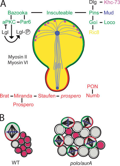
Over the past decade, many of the key components of the genetic machinery that regulate the asymmetric division of Drosophila melanogaster neural progenitors, neuroblasts, have been identified and their functions elucidated. Studies over the past two years have shown that many of these identified components act to regulate the self-renewal versus differentiation decision and appear to function as tumor suppressors during larval nervous system development. In this paper, we highlight the growing number of molecules that are normally considered to be key regulators of cell cycle events/progression that have recently been shown to impinge on the neuroblast asymmetric division machinery to control asymmetric protein localization and/or the decision to self-renew or differentiate.


Figures

Figure 1.
Figure 1.
Summary of some of the key players and features of neuroblast asymmetric division. (A) Asymmetrical segregation of basal cell fate determinants specifically into the daughter GMC requires the correct localization of protein complexes to the apical cell cortex. The apically localized proteins comprise two protein complexes linked by the adaptor protein Inscuteable. The evolutionary conserved Par protein cassette comprising Bazooka/Par3, aPKC, and Par6 is the first protein complex to localize to the neuroblast cell cortex and is primarily involved in excluding the basally localized proteins from the apical cortex. This protein cassette regulates the activity of the tumor suppressor lethal giant larvae (Lgl), which is also essential for correct targeting of the basal protein complexes. Par6 can directly associate with Lgl, and it is in this complex that aPKC is believed to inactivate Lgl by phosphorylation. Miranda is thus recruited to the basal cell cortex by the active nonphosphorylated Lgl. The second apical protein complex contains proteins involved with heterotrimeric G protein signaling, including Gαi, Partner of Inscuteable (Pins), and Locomotion defects (Loco). This complex is thought to mediate a receptor-independent heterotrimeric G protein signaling mechanism involving the regulation of Gαi through interactions with the cytoplasmic guanine nucleotide exchange factor (Ric-8) and guanine nucleotide dissociation inhibitors (Loco and Pins). The Gαi–Pins–Loco complex mediates mitotic spindle formation and alignment to ensure that the cleavage plane is orthogonal to the apical/basal polarity axis. The geometry of the neuroblast mitotic spindle is asymmetrical; the spindle length is longer on the apical side, and the entire spindle is displaced toward the basal cortex. The centrosomes are also nonequivalent, with the larger mother centrosome emanating more extensive astral microtubules and being preferentially retained within the neuroblast through subsequent divisions. Pins can also associate with the centrosome- and apical cortex–associated nuclear mitotic apparatus protein–-related protein mushroom body defective (Mud), which is essential for proper spindle alignment, as well as Discs large (Dlg) and the astral microtubule plus end protein Khc-73 to induce cortical polarity. The actin/myosin cytoskeleton also plays an important role in the assembly of these apical/basal protein complexes. Actin filaments but not microtubules appear to play an essential role in cortical tethering of the proteins, and the Drosophila myosins II (Zipper) and VI (Jaguar) exist in mutually exclusive complexes with Miranda and are essential for correct asymmetric localization of the cell fate determinants. The basal proteins exist as two protein complexes. One complex contains the adaptor protein Miranda, which associates with and facilitates the asymmetric localization of the translational repressor Brain tumor (Brat), the homeodomain transcription factor Prospero, and the double-stranded RNA-binding protein Staufen, which itself can bind prospero transcripts. The second complex contains the Notch antagonist Numb and its binding partner Partner of Numb (Pon). Upon segregation into the GMC, Miranda is degraded, allowing Prospero to translocate into the nucleus to activate genes involved in differentiation and repress genes involved in proliferation. The GMC divides terminally to produce two neurons or glia. Note that the apical/basal nomenclature is based on embryonic neuroblasts and that neuroblasts in the central brain divide without a fixed orientation. Please note that the color of the lettering corresponds to the protein's localization in the schematic picture; in the case of black lettering, the protein can be found throughout the cortex. (B) Postembryonic neuroblasts divide to produce a lineage of differentiated progeny. The cell types of the lineage can readily be distinguished with neuroblast markers such as Insc (green), Miranda (red), and Deadpan (gray) and markers for differentiated progeny like Elav and nuclear Prospero (red). A disruption to cell polarity and/or spindle orientation (e.g., in aurora A and polo mutants) can affect the balance between self-renewal and differentiation, resulting in too many self-renewing cells at the expense of differentiated progeny. WT, wild type.

Comment in

Similar articles

Cited by

References

    1. Almeida, M.S., and S.J. Bray. 2005. Regulation of post-embryonic neuroblasts by Drosophila Grainyhead. Mech. Dev. 122:1282–1293. - PubMed
    1. Ando, K., T. Ozaki, H. Yamamoto, K. Furuya, M. Hosoda, S. Hayashi, M. Fukuzawa, and A. Nakagawara. 2004. Polo-like kinase 1 (Plk1) inhibits p53 function by physical interaction and phosphorylation. J. Biol. Chem. 279:25549–25561. - PubMed
    1. Audibert, A., F. Simon, and M. Gho. 2005. Cell cycle diversity involves differential regulation of Cyclin E activity in the Drosophila bristle cell lineage. Development. 132:2287–2297. - PubMed
    1. Beaucher, M., E. Hersperger, A. Page-McCaw, and A. Shearn. 2007. Metastatic ability of Drosophila tumors depends on MMP activity. Dev. Biol. 303:625–634. - PubMed
    1. Bello, B., H. Reichert, and F. Hirth. 2006. The brain tumor gene negatively regulates neural progenitor cell proliferation in the larval central brain of Drosophila. Development. 133:2639–2648. - PubMed

Publication types

Substances

LinkOut - more resources