

Nondestructive imaging of live human keloid and facial tissue using multiphoton microscopy
- PMID: 18209122
- PMCID: PMC4144461
- DOI: 10.1001/archfacial.2007.18
Nondestructive imaging of live human keloid and facial tissue using multiphoton microscopy
Abstract
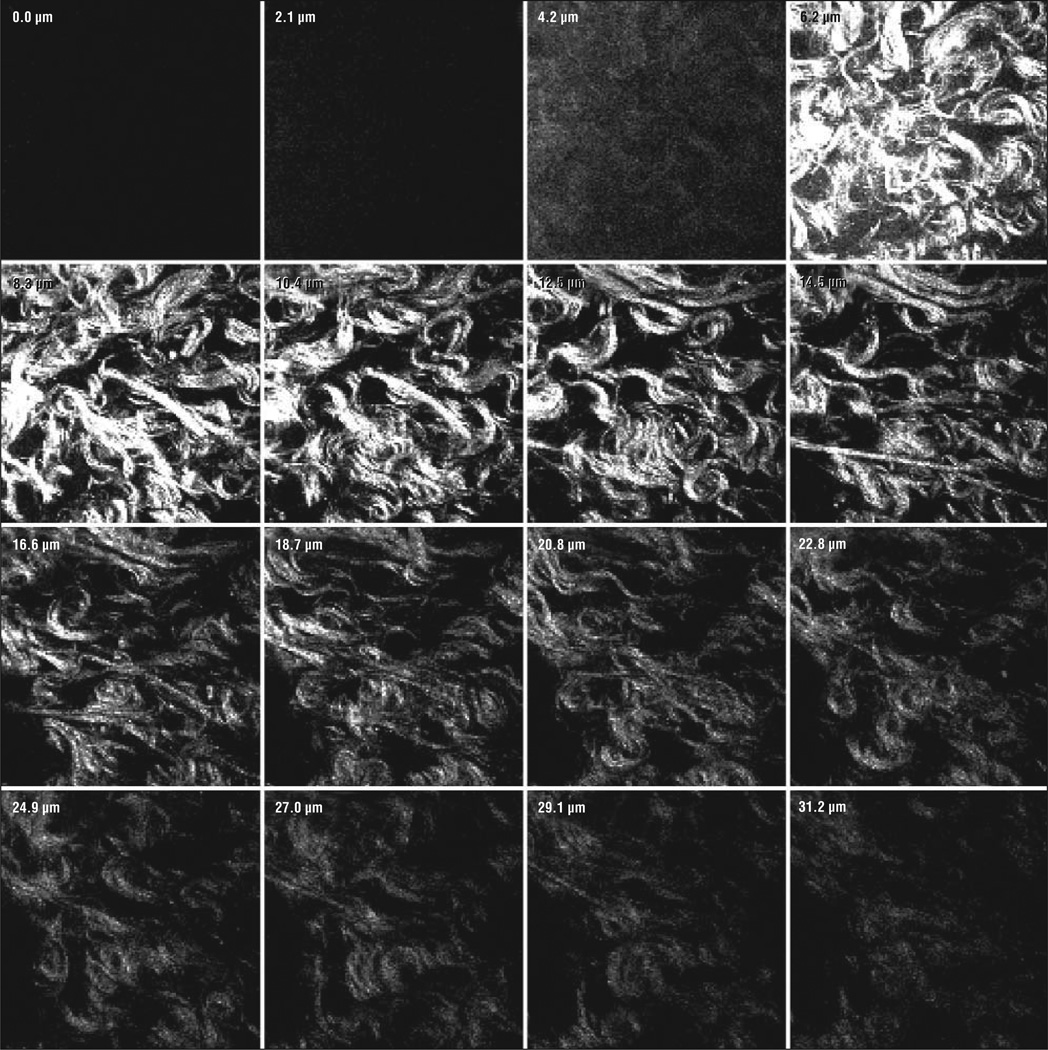
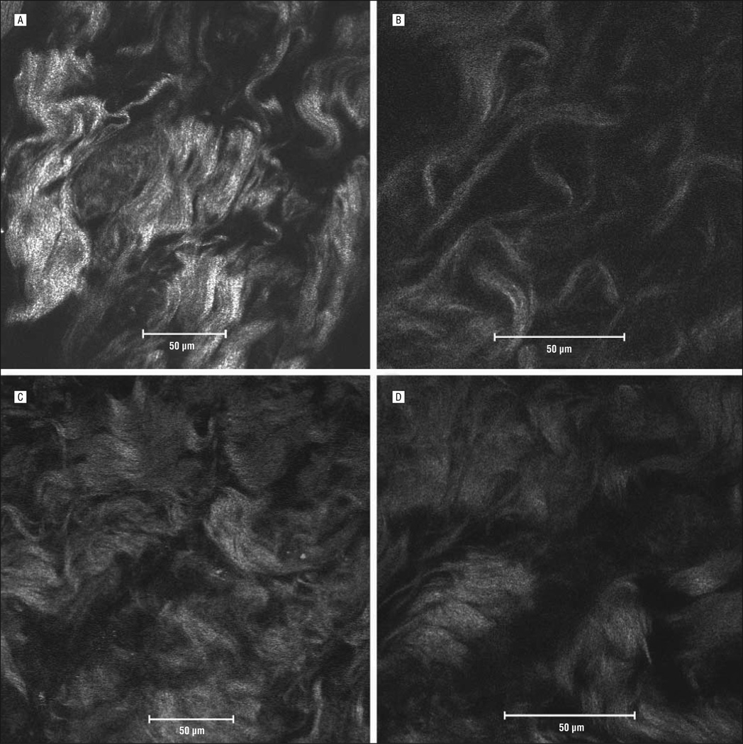
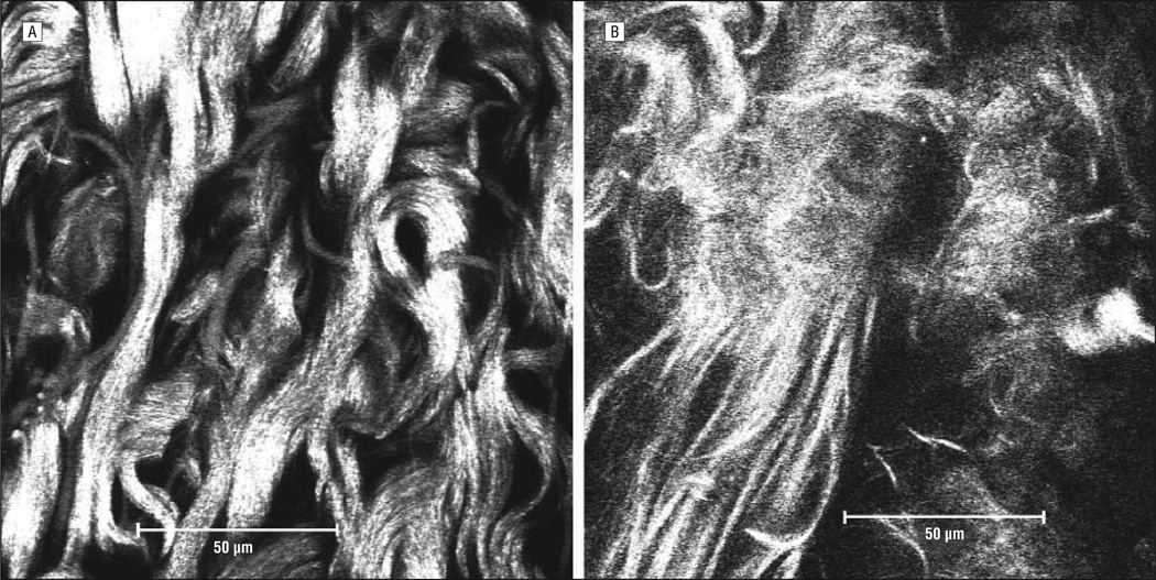
Objectives: To use multiphoton microscopy to image collagen fibers and matrix structure in nonfixed human keloid tissue and normal human facial skin obtained following surgery and to compare the findings to existing knowledge of normal skin and keloid morphology to determine if this technology is a suitable adjunct for conventional histology.
Methods: Epidermis was removed to expose the fibroblast-rich dermal layer that was then imaged using a multiphoton confocal microscope (Zeiss-Meta 510; Carl Zeiss, Jena, Germany). An 800-nm tunable titanium/sapphire femtosecond laser (Mai-Tai; Newport Co Spectra-Physics, Mountain View, California) was used to excite the tissue; second harmonic generation between 397 and 408 nm and autofluorescent signals were collected. Images were obtained using a Plan-Neofluar x40 oil immersion objective lens and a Plan-Apochromat x63 oil immersion lens.
Results: Compared with normal skin, keloids showed disorganized collagen fibers arranged in complex swirls and bundles 20 to 30 microm in diameter. Normal tissue showed collagen fibers as distinct, straight strands less than 10 microm in diameter. Differences between normal and keloid tissue were subtle but apparent.
Conclusions: The value of imaging living tissue is a significant benefit. Because keloids and hypertrophic scars result from altered collagen metabolism, the development of clinical multiphoton microscopy systems may allow examination of wound healing dynamics in vivo and potentially provides a means to monitor therapy without the need for biopsy or the risk of injury to tissue.
Figures
References
-
- Al-Attar A, Mess S, Thomassen JM, Kauffman CL, Davison SP. Keloid pathogenesis and treatment. Plast Reconstr Surg. 2006;117(1):286–300. - PubMed
-
- Marneros AG, Norris JE, Olsen BR, Reichenberger E. Clinical genetics of familial keloids. Arch Dermatol. 2001;137(11):1429–1434. - PubMed
-
- Navarro FA, So PT, Nirmalan R, et al. Two-photon confocal microscopy: a nondestructive method for studying wound healing. Plast Reconstr Surg. 2004;114(1):121–128. - PubMed
-
- Emptage NJ. Fluorescent imaging in living systems. Curr Opin Pharmacol. 2001;1(5):521–525. - PubMed
-
- White N, Errington R. Multi-photon microscopy: seeing more by imaging less. Biotechniques. 2002;33(2):298–300. 302, 304–305. - PubMed
Publication types
MeSH terms
Grants and funding
LinkOut - more resources
Full Text Sources
Other Literature Sources