

Muscle study in experimental scoliosis in rabbits with costotransversectomy: evidence of ischemic process
- PMID: 18210168
- PMCID: PMC2367410
- DOI: 10.1007/s00586-008-0598-9
Muscle study in experimental scoliosis in rabbits with costotransversectomy: evidence of ischemic process
Abstract
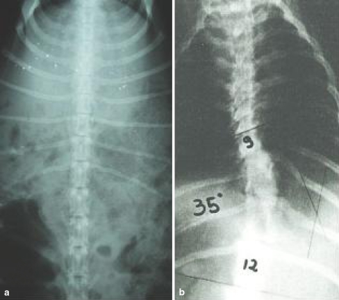
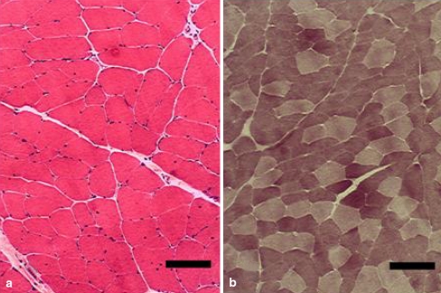
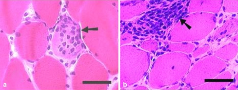
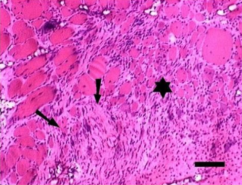
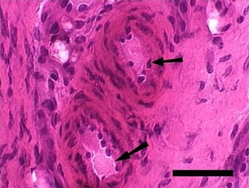
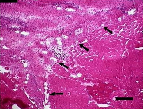
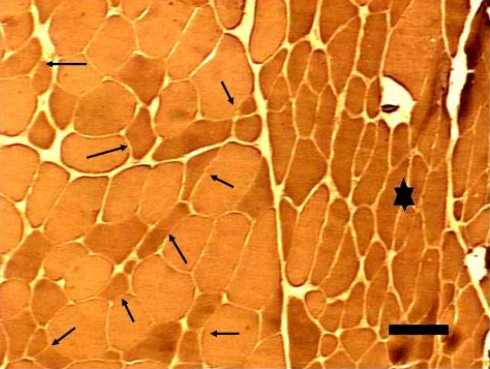
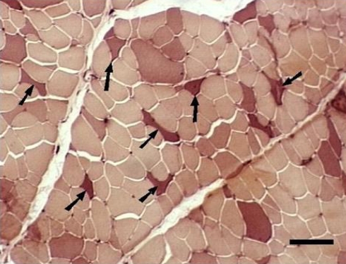
Scoliosis involves the central nervous system diseases, ligaments, articulations and skeletal muscles, but there is no consensus on its pathogeny and progression of muscle abnormalities. In this study, we investigate the morphologic changes in the muscle of rabbit submitted to experimental scoliosis, with special emphasis on abnormalities related to blood supply. We studied 26 rabbits subjected to costotransversectomy by pulling out six transverse processes at thoracic level and six rabbits were used as controls. All the animals operated upon developed scoliosis showing an average angle of 29.1 degrees on the 60th day, with its apices located at T4 and T12 when they were subjected to paraspinal muscle biopsy on both sides. The muscle biopsies were subjected to histological stains and histochemical reactions, as well as to a morphometric study. On the concave side, the changes were not statistically significant regarding the control group. On the convex side conjunctive tissue proliferation, infiltration by adipose tissue, central nucleus excess, inflammatory reaction, segmental fibrosis, type 1 fiber hypertrophy, type 2 fiber hypertrophy and atrophic angular dark fibers in the unspecific esterase were statistically significant. The segmental fibrosis reached a circumscribed muscle segment, compatible with an ischemic phenomenon. The histological diagnoses on the concave side of the animals had unspecific alterations (atrophy and hypertrophy) in 13, myopathy in 3, denervation in 3 and normal in 7. The convex side diagnoses were myopathy in 14, denervation in 8, mixed in 3 and normal in 1. The procedure determined morphologic changes on the convex side indicating possible denervation or myopathy of ischemic origin.
Figures
Similar articles
-
Paraspinal muscle pathology in experimental scoliosis.Arch Orthop Trauma Surg. 1989;108(6):342-5. doi: 10.1007/BF00932442. Arch Orthop Trauma Surg. 1989. PMID: 2619518
-
[Histochemical changes of muscle fibers and motor end-plates of paravertebral muscles in scoliosis associated with syringomyelia].Zhongguo Yi Xue Ke Xue Yuan Xue Bao. 2006 Dec;28(6):790-4. Zhongguo Yi Xue Ke Xue Yuan Xue Bao. 2006. PMID: 17260468 Chinese.
-
Paraspinal muscle fibre type alterations associated with scoliosis: an old problem revisited with new evidence.Eur Spine J. 1998;7(4):289-93. doi: 10.1007/s005860050077. Eur Spine J. 1998. PMID: 9765036 Free PMC article.
-
Histochemical analysis of paraspinal rotator muscles from patients with adolescent idiopathic scoliosis: a cross-sectional study.Medicine (Baltimore). 2015 Feb;94(8):e598. doi: 10.1097/MD.0000000000000598. Medicine (Baltimore). 2015. PMID: 25715269 Free PMC article.
-
[Radiological comparison of bilateral paravertebral muscles in degenerative lumbar scoliosis and its potential importance].Zhonghua Wai Ke Za Zhi. 2012 Nov;50(11):975-80. Zhonghua Wai Ke Za Zhi. 2012. PMID: 23302479 Chinese.
Cited by
-
Effect of neurocentral cartilage destruction on spinal growth in immature rabbits.J Int Med Res. 2019 Feb;47(2):951-961. doi: 10.1177/0300060518820198. Epub 2019 Jan 7. J Int Med Res. 2019. PMID: 30616424 Free PMC article.
-
Chronic Paraspinal Muscle Injury Model in Rat.J Korean Neurosurg Soc. 2016 Sep;59(5):430-6. doi: 10.3340/jkns.2016.59.5.430. Epub 2016 Sep 8. J Korean Neurosurg Soc. 2016. PMID: 27651859 Free PMC article.
References
-
- Ahn UM, Ahn NU, Nallamshetty L, Buchowski JM, Rose PS, Miller NH, Kostuik JP, Sponseller PD. The etiology of adolescent idiopathic scoliosis. Am J Orthop. 2002;31:387–395. - PubMed
-
- Banker BQ, Engel AG. Basic reaction of muscle. In: Engel AG, Franzini-Armstrong C, editors. Myology, basic and clinical. New York: McGraw-Hill; 1994. pp. 832–888.
MeSH terms
LinkOut - more resources
Full Text Sources
Medical
