. 2008 Feb;41(1):136-55.
doi: 10.1111/j.1365-2184.2007.00500.x.

Identification of genes deregulated during serum-free medium adaptation of a Burkitt's lymphoma cell line

Affiliations

Identification of genes deregulated during serum-free medium adaptation of a Burkitt's lymphoma cell line

L Zander et al. Cell Prolif. 2008 Feb.

Abstract

Objective: Serum is usually added to growth media when mammalian cells are cultured in vitro to supply the cells with growth factors, hormones, nutrients and trace elements. Defined proteins and metal ions, such as insulin, growth factors, transferrin and sodium selenite, are sometimes also included and can in some cases substitute serum components. How adaptation to serum free media influences cells has not been studied in detail.

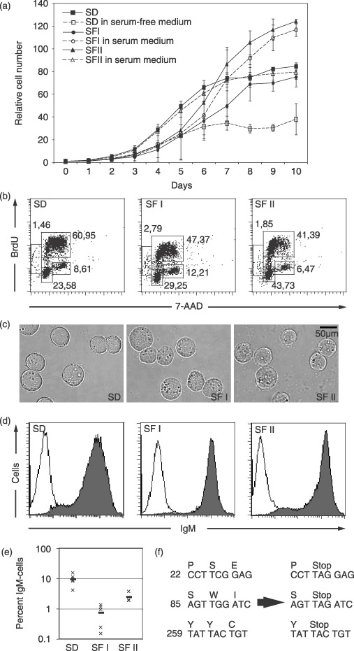
Materials and methods: We have adapted the Burkitt's lymphoma line Ramos to a serum-free medium that supports long-term survival and studied gene expression changes that occurred during the adaptation process.

Results and conclusions: The adaptation process was characterized by initial cell population growth arrest, and after that extensive cell death, followed by proliferation and long-term survival of clonal cultures. Proliferation and cell cycle progression of the serum-free cultures closely mimicked that of serum-dependent cells. Affymetrix micro-array technology was used to identify gene expression alterations that had occurred during the adaptation. Most changes were subtle, but frequently the genes with altered expression were involved in basal cellular functions such as cell division, cell cycle regulation, apoptosis and cell signalling. Some alterations were restored when the cells were transferred back to serum-containing medium, indicating that expression of these genes was controlled by components in serum. Others were not, and may represent changes that were selected during the adaptation process. Among these were, for example, several genes within the Wnt signalling pathway.


Figures

Figure 1
Figure 1
Phenotype of serum‐dependent Ramos cells and the serum‐free cultures SFI and SFII. (a) Cells were cultured in the indicated media and were counted for 10 days to determine proliferation rates. Serum‐dependent cells in normal serum‐containing medium (filled squares) and serum‐free medium (open squares), SFI and SFII cells cultured in serum‐free medium (filled circles and filled triangles, respectively) or serum‐containing medium (open circles and open triangles). Average of three experiments is presented together with the highest and lowest values shown as error bars. (b) Cell cycle progression of serum‐dependent Ramos cells and serum‐free SFI and SFII cultures, using BrdU and 7‐AAD labelling are shown. Relative sizes for the major populations (G1, S and G2) are shown from a representative experiment. (c) Photographs of serum‐dependent Ramos cells and the serum‐free cultures (d) IgM surface expression of serum‐dependent Ramos cells and the SFI and SFII serum‐free cultures. (e) Percentage of cells that not expressing IgM 7 weeks after subcloning are shown. For SD six distinct clones are shown, for SFI five distinct clones are shown and for SFII three clones are shown. Thick line indicates average number of non‐expressors. (f) Stop codons identified in IgM‐negative cells generated during culture of subclones. IgM non‐expressing cells were sorted from the subclone cultures, DNA prepared and the antibody heavy chain gene sequenced. Three examples of stop codon identified are shown.
Figure 2
Figure 2
Scatter plots of micro‐array results. (a) Mean intensity value of serum‐dependent cells plotted against mean intensity value of serum‐free cells. Genes classified as absent are marked in grey and genes further analysed in black. Some significantly deregulated genes are marked. (b) Scatter plot of mean intensity values of randomized groups (see Material and Methods).
Figure 3
Figure 3
Micro‐array results were validated using Northern blot and FACS analysis. (a) Northern blot analyses of representative genes up‐regulated in serum‐free cells. Data from the Northern blot analysis are black bars and from micro‐array analysis are grey bars. Data are presented as change in expression compared to serum‐dependent cells. (b) Northern blot analyses of representative genes down‐regulated in serum‐free cells. (c) FACS analysis of HLA‐DR expression of SD cells, SFI, SFII and SFI cells re‐cultured in serum medium for 2 days. (d) Changes that occurred only in one of the cultures are validated using Northern blotting.
Figure 4
Figure 4
Changes in gene expression that occurred when serum‐free cells were re‐transferred to serum containing medium, validated using real‐time PCR or Northern blots. (a) Real‐time PCR assays of genes of the Wnt pathway. (b,c) Expression of LOC51315 and SMAD1 in SFI cells when re‐transferred to serum containing medium for 2 h, 8 h and 2 days using northern blot. (d,e) Northern blot analysis of genes differentially expressed between SFI and SFII cultures. RNA from SD, SFI, SFII cells re‐cultured in serum medium for 2 h, 8 h and 2 days, SFI and SFII cells re‐cultured in medium containing serum for 2 days were used. Data are presented as changes in expression compared to serum‐dependent cells.

References

    1. Abramovich C, Shen WF, Pineault N, Imren S, Montpetit B, Largman C, Humphries RK (2000) Functional cloning and characterization of a novel nonhomeodomain protein that inhibits the binding of PBX1‐HOX complexes to DNA. J Biol Chem 275, 26172–26177. - PubMed
    1. Badea T, Niculescu F, Soane L, Fosbrink M, Sorana H, Rus V, Shin ML, Rus H (2002) RGC‐32 increases p34CDC2 kinase activity and entry of aortic smooth muscle cells into S‐phase. J Biol Chem 277, 502–508. - PubMed
    1. Baglia LA, Cruz D, Shaw JE (1991) An Epstein‐Barr virus‐negative Burkitt lymphoma cell line (sfRamos) secretes a prolactin‐like protein during continuous growth in serum‐free medium. Endocrinology 128, 2266–2272. - PubMed
    1. Baglia LA, Cruz D, Shaw JE (1992) Production of immunoreactive forms of growth hormone by the Burkitt tumor serum‐free cell line sfRamos. Endocrinology 130, 2446–2454. - PubMed
    1. Barker N, Clevers H (2000) Catenins, Wnt signaling and cancer. Bioessays 22, 961–965. - PubMed

Publication types

Substances