. 2008 Apr;28(7):2314-23.
doi: 10.1128/MCB.00998-07. Epub 2008 Jan 22.

Trio mediates netrin-1-induced Rac1 activation in axon outgrowth and guidance

Affiliations

Trio mediates netrin-1-induced Rac1 activation in axon outgrowth and guidance

Anne Briançon-Marjollet et al. Mol Cell Biol. 2008 Apr.

Abstract

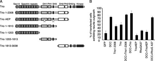
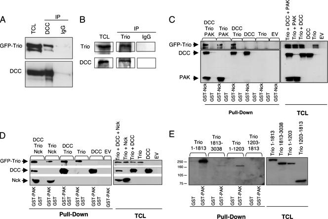
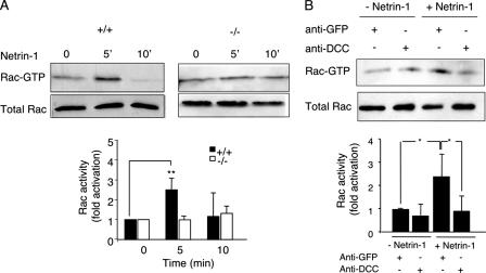
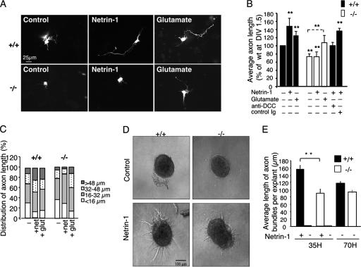
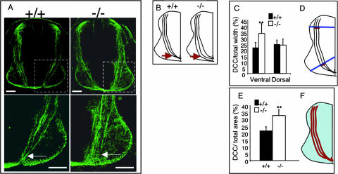
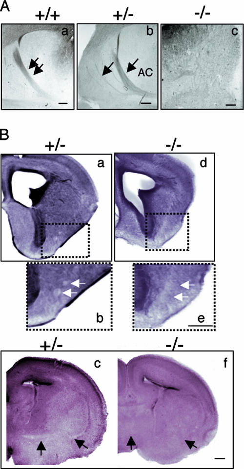
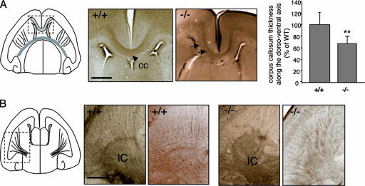
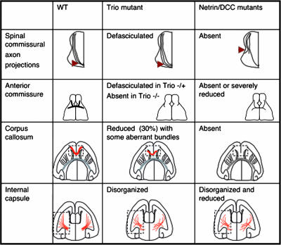
The chemotropic guidance cue netrin-1 promotes neurite outgrowth through its receptor Deleted in Colorectal Cancer (DCC) via activation of Rac1. The guanine nucleotide exchange factor (GEF) linking netrin-1/DCC to Rac1 activation has not yet been identified. Here, we show that the RhoGEF Trio mediates Rac1 activation in netrin-1 signaling. We found that Trio interacts with the netrin-1 receptor DCC in mouse embryonic brains and that netrin-1-induced Rac1 activation in brain is impaired in the absence of Trio. Trio(-/-) cortical neurons fail to extend neurites in response to netrin-1, while they are able to respond to glutamate. Accordingly, netrin-1-induced commissural axon outgrowth is reduced in Trio(-/-) spinal cord explants, and the guidance of commissural axons toward the floor plate is affected by the absence of Trio. The anterior commissure is absent in Trio-null embryos, and netrin-1/DCC-dependent axonal projections that form the internal capsule and the corpus callosum are defective in the mutants. Taken together, these findings establish Trio as a GEF that mediates netrin-1 signaling in axon outgrowth and guidance through its ability to activate Rac1.


Figures

FIG. 1.
FIG. 1.
Trio defective in Rac1 activation inhibits DCC-induced neurite outgrowth in N1E-115 neuroblastoma cells. (A) Schematic of Trio and Trio mutant proteins. TrioAEP is a dominant negative form of Trio and corresponds to Trio(1-2308) containing triple mutations (stars) in the GEFD1 and adjacent SH3 domain. DH, Dbl homology; PH, pleckstrin homology. (B) N1E-115 cells were transfected with the indicated plasmids, and cells exhibiting neurite outgrowth were counted 24 h after transfection. The values correspond to the average of at least three independent experiments. Error bars represent standard deviations.
FIG. 2.
FIG. 2.
Interaction of DCC and Trio. (A) Lysates of HEK-293 cells transfected with pEGFP-Trio and pRK5-DCC were submitted to immunoprecipitation using anti-DCC antibodies (DCC) or mouse IgG coupled to protein G-Sepharose beads. Immunoprecipitated proteins and 10% of the total cell lysates were submitted to SDS-PAGE, and GFP-Trio and DCC were detected by Western blotting using anti-DCC and anti-GFP antibodies. (B) Lysates of E18.5 mouse brains were submitted to immunoprecipitation using anti-Trio or normal rabbit IgGs coupled to protein G-Sepharose beads. Immunoprecipitated proteins and 10% of the total cell lysates were submitted to SDS-PAGE, and the presence of DCC and Trio was detected by Western blotting using the appropriate antibodies. (C and D) Lysates of COS-7 cells transfected with pRK5 (EV), pRK5-DCC, pEGFP-Trio, pRK5-HA-Nck-1, or pRK5myc-PAK1, alone or as indicated, were incubated with GST and with GST-Nck (C) or GST-PAK (D). GST pull-down proteins (pull-down) and 10% of the total cell lysates were submitted to SDS-PAGE, and proteins were detected by Western blotting analysis using anti-DCC, anti-GFP, anti-PAK1, and anti-Nck-1 antibodies. (E) Lysates of COS-7 cells transfected with pEGFP-Trio(1-1813), pEGFP-Trio(1813-3038), pEGFP-Trio(1-1203), or pEGFP-Trio(1203-1813) were incubated with GST or GST-PAK. GST pull-down proteins (pull-down) and 8% of the total cell lysates were submitted to SDS-PAGE, and proteins were detected by Western blotting analysis using anti-GFP antibodies. IP, immunoprecipitation; TCL, total cell lysates.
FIG. 3.
FIG. 3.
Netrin-1-induced Rac1 activation is impaired in Trio null-embryonic brains. (A) GTP-loaded Rac1 was pulled down using GST-PAK-PBD from lysates of Trio+/+ or Trio−/− embryonic brains treated or not with netrin-1 for the indicated time. GTP-bound Rac1 was detected by Western blotting using anti-Rac1 antibodies (top). Total cell lysates probed for Rac1 indicated equal amounts of GTPase (bottom). Quantification of Rac1 activity corresponds to the average of at least three independent experiments (P < 0.01). (B) The experiment was as described in panel A, except that DCC blocking antibodies (4 μg/ml) were added before netrin-1 stimulation (5 min). Mouse anti-GFP antibodies were used as a negative control and had no effect on netrin-1-induced Rac1 activation. Quantification of Rac1 activity corresponds to the average of at least three independent experiments (P = 0.012, Student t test). For both panels, error bars represent standard deviations.
FIG. 4.
FIG. 4.
Trio is required for netrin-1-induced axon outgrowth. (A) Neurite outgrowth of Trio+/+ or Trio−/− cortical neurons expressing GFP at day in vitro 1.5 treated with control buffer, netrin-1, or glutamate for 24 h. Scale bar, 25 μm. (B) Quantification of average axon length of cortical neurons presented in panel A. Values are represented as a percentage of average axon length of wt cortical neurons at day in vitro (DIV) 1.5 incubated with control buffer. When indicated, neurons were incubated with control Igs or DCC blocking antibodies before netrin-1 addition. **, P <0.001 for the comparison to wt neurons expressing GFP, except for the dotted line that refers to GFP-transfected Trio-null neurons (n = 8 for +/+ and n = 10 for −/− embryos). (C) Distribution of axon length from panel B. (D) E11.5 dorsal spinal cord explants from Trio+/+ or Trio−/− embryos were incubated with control buffer or netrin-1 for 35 h. Scale bar, 100 μm. (E) Quantification of the average length of axon bundles per explant after a 35-h incubation with netrin-1 (n = 10 for Trio+/+, and n = 4 for Trio−/− embryos; **, P < 0.001) or after 70 h in the absence of netrin-1 (n = 3 for +/+ and −/− embryos).
FIG. 5.
FIG. 5.
Commissural axon projections are defective in Trio-null embryos. (A) In the upper frames, trajectories of commissural axons are visualized using anti-DCC antibodies in sections of Trio+/+ or Trio−/− E11.5 embryos. The lower frames show enlargements of the corresponding images. Scale bar, 80 μm. (B) On the left is a schematic representing normal commissural axons that project from the dorsal spinal cord toward the ventral floor plate. In Trio−/− embryos (right), commissural axons are defasciculated when they reach the ventral floor plate (arrows in both A and B). (C and D) The thickness of axon bundles in the dorsal and ventral spinal cords was quantified by measuring the width of the DCC-stained axons (red) relative to the width of the spinal cord (blue) as depicted in panel D. (E and F) Axon defasciculation in Trio−/− embryos was quantified by measuring the DCC-stained area (red) relative to the total area of the spinal cord (blue) as depicted in panel F. P < 0.001, Student t test. Error bars represent standard deviations (n = 5 for +/+ and n = 7 for −/− embryos).
FIG. 6.
FIG. 6.
The anterior commissure is absent in Trio-null embryos. (A) Neuropilin 2 (Nrp2) immunostaining on horizontal serial brain sections from E17 +/+ (a), +/− (b), or −/− (c) embryos (n = 6 for +/+ and −/− embryos; n = 4 for +/− embryos). In the heterozygous embryos, the anterior branch of the commissure (AC) is defasciculated, which is illustrated by several roots exiting the cortex at lateral positions (arrows in frame b). In the Trio-null embryos, the commissure is absent (c). Scale bar, 80 μm. (B) Eosin staining on coronal brain sections from E17 +/− (a, b, and c) and −/− (d, e, and f) embryos. Defasciculated fibers are present in the heterozygous embryos (b, white arrows), whereas they are absent in the Trio-null embryos (e). In more posterior sections, anterior commissural fibers are detected in the heterozygous (c, black arrows) but not in Trio-null brains (f). Scale bar, 300 μm.
FIG. 7.
FIG. 7.
Defects in the corpus callosum and internal capsule in Trio-null embryos. (A) DCC immunostaining on horizontal brain sections from E18.5 Trio+/+ and Trio−/− embryos showing the corpus callosum (CC) region. In the Trio-null embryos, the corpus callosum appears slightly abnormal with some defasciculated fibers (arrows) in the horizontal sections. The right panel represents the quantification of corpus callosum thickness along the dorsoventral axis in Trio+/+ and Trio−/− embryos. Quantification has been obtained by counting the number of horizontal sections in which the corpus callosum is present and dividing by the total number of sections. The corpus callosum thickness of the Trio-null embryos is expressed relative to the thickness of the wt corpus callosum along the dorsoventral axis (n = 5 for Trio+/+ embryos; n = 8 for Trio−/− embryos; P < 0.05). Scale bar, 50 μm. (B) DCC immunostaining on horizontal brain sections from E18.5 Trio+/+ and Trio−/− embryos showing the internal capsule (IC) region. DCC-positive fibers are clearly disorganized in the internal capsule of Trio−/− embryos. Two different examples are shown. This defect was observed in 8 out of 9 Trio−/− embryos. Scale bar, 50 μm.
FIG. 8.
FIG. 8.
Comparison of the phenotypes observed in the Trio-null embryos with netrin-1- and DCC-deficient embryos. Four different netrin-1- and DCC-dependent neuronal projections were examined in the Trio-null embryos, namely, the spinal commissural axon projections, the anterior commissure, the corpus callosum, and the internal capsule. The figure shows the comparison between the phenotypes observed in Trio-, netrin-1-, and DCC-deficient embryos (7, 13, 43).

References

    1. Ackerman, S. L., L. P. Kozak, S. A. Przyborski, L. A. Rund, B. B. Boyer, and B. B. Knowles. 1997. The mouse rostral cerebellar malformation gene encodes an UNC-5-like protein. Nature 386838-842. - PubMed
    1. Awasaki, T., M. Saito, M. Sone, E. Suzuki, R. Sakai, K. Ito, and C. Hama. 2000. The Drosophila trio plays an essential role in patterning of axons by regulating their directional extension. Neuron 26119-131. - PubMed
    1. Barallobre, M. J., M. Pascual, J. A. Del Rio, and E. Soriano. 2005. The Netrin family of guidance factors: emphasis on Netrin-1 signalling. Brain Res. Brain Res. Rev. 4922-47. - PubMed
    1. Bateman, J., H. Shu, and D. Van Vactor. 2000. The guanine nucleotide exchange factor Trio mediates axonal development in the Drosophila embryo. Neuron 2693-106. - PubMed
    1. Bellanger, J. M., J. B. Lazaro, S. Diriong, A. Fernandez, N. Lamb, and A. Debant. 1998. The two guanine nucleotide exchange factor domains of Trio link the Rac1 and the RhoA pathways in vivo. Oncogene 16147-152. - PubMed

Publication types

MeSH terms

Substances