. 2008 Apr;28(7):2368-79.
doi: 10.1128/MCB.01780-07. Epub 2008 Jan 22.

Wnt-3a and Dickkopf-1 stimulate neurite outgrowth in Ewing tumor cells via a Frizzled3- and c-Jun N-terminal kinase-dependent mechanism

Affiliations

Wnt-3a and Dickkopf-1 stimulate neurite outgrowth in Ewing tumor cells via a Frizzled3- and c-Jun N-terminal kinase-dependent mechanism

Yoshimi Endo et al. Mol Cell Biol. 2008 Apr.

Abstract

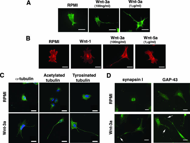
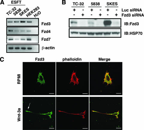
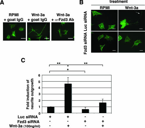
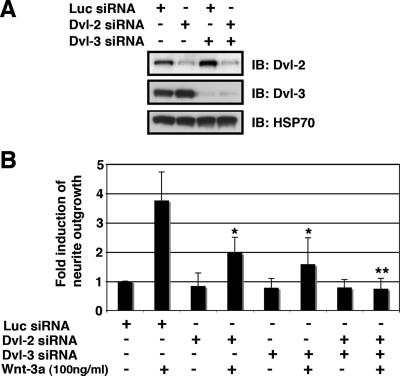
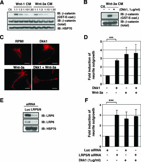
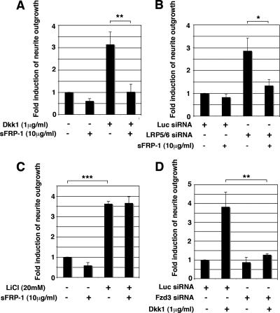
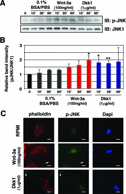
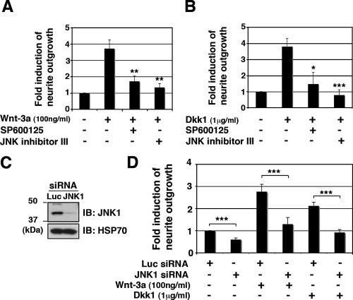
Recombinant Wnt-3a stimulated the rapid formation of elongated processes in Ewing sarcoma family tumor (ESFT) cells that were identified as neurites. The processes stained positively for polymerized actin and microtubules as well as synapsin I and growth-associated protein 43. Inhibition of the Wnt receptor, Frizzled3 (Fzd3), with antiserum or by short interfering RNA (siRNA) markedly reduced neurite extension. Knockdown of Dishevelled-2 (Dvl-2) and Dvl-3 also suppressed neurite outgrowth. Surprisingly, disruption of the Wnt/Fzd/lipoprotein receptor-related protein (LRP) complex and the associated beta-catenin signaling by treating cells either with the Wnt antagonist Dickkopf-1 (Dkk1) or LRP5/LRP6 siRNA enhanced neuritogenesis. Neurite outgrowth induced by Dkk1 or with LRP5/LRP6 siRNA was inhibited by secreted Fzd-related protein 1, a Wnt antagonist that binds directly to Wnt. Moreover, Dkk1 stimulation of neurite outgrowth was blocked by Fzd3 siRNA. These results suggested that Dkk1 shifted endogenous Wnt activity from the beta-catenin pathway to Fzd3-mediated, noncanonical signaling that is responsible for neurite formation. In particular, c-Jun amino-terminal kinase (JNK) was important for neurite outgrowth stimulated by both Wnt-3a and Dkk1. Our data demonstrate that Fzd3, Dvl, and JNK activity mediate Wnt-dependent neurite outgrowth and that ESFT cell lines will be useful experimental models for the study of Wnt-dependent neurite extension.


Figures

FIG. 1.
FIG. 1.
Wnt-3a stimulated neurite outgrowth in ESFT cell lines. TC-32 cells were seeded on glass coverslips precoated with collagen I and subsequently treated for 3 h with serum-free RPMI medium in the presence or absence of recombinant Wnt-3a. (A) Alexa Fluor 488 phalloidin staining of representative cells from cultures incubated with serum-free RPMI alone (control) or supplemented with Wnt-3a (100 ng/ml or 1 μg/ml). Scale bars, 15 μm. (B) TC-32 cell morphology following treatment with different Wnt ligands. Alexa Fluor 568 phalloidin staining of representative cells from cultures incubated with RPMI (control), partially purified Wnt-1, Wnt-3a (100 ng/ml), or Wnt-5a (1 μg/ml). Scale bars, 20 μm. (C) Staining patterns of anti-α-tubulin, acetylated tubulin, and tyrosinated tubulin in TC-32 cells treated with RPMI control or Wnt-3a (100 ng/ml). Tubulin antibodies stained green; blue (DAPI) stain highlights nuclei. Scale bars, 20 μm. (D) TC-32 cells treated with RPMI medium alone or with Wnt-3a (100 ng/ml) were stained with synapsin I or GAP-43 antibodies. Arrows indicate Wnt-3a-induced neurite outgrowth. Scale bars, 20 μm.
FIG. 2.
FIG. 2.
Fzd3 is expressed in ESFT cell lines. (A) RT-PCR analysis of Fzd expression in three ESFT cell lines. cDNA from HEK293 cells was used as a positive control for Fzd expression. β-Actin RT-PCR was performed as an internal control of RNA analysis. (B) Immunoblot (IB) analysis of Fzd3 protein in three ESFT lines after siRNA treatment. Cells were transfected with either Fzd3 or Luc siRNA, and protein levels were determined 48 h after transfection with Fzd3 antibody raised against a carboxy-terminal peptide of Fzd3. Immunoblotting with anti-HSP70 was performed as a loading control. −, absence of; +, presence of. (C) Immunostaining of Fzd3 in TC-32 cells. TC-32 cells were treated with serum-free RPMI in the presence or absence of Wnt-3a (100 ng/ml) for 3 h and stained with anti-Fzd3 antibody and Alexa Fluor 568 phalloidin. Note prominent Fzd3 staining at the tip of the neurite (arrow). Scale bars, 20 μm.
FIG. 3.
FIG. 3.
Fzd3 mediated Wnt-3a-dependent neurite outgrowth. (A) TC-32 cells were pretreated with either antibody against the amino-terminal, extracellular domain of mouse Fzd3 or control goat IgG, followed by incubation with or without Wnt-3a (100 ng/ml) for 3 h. Cell morphology was visualized by Alexa Fluor 488 phalloidin staining. Scale bars, 20 μm. (B) TC-32 cells were transfected with either Fzd3 or Luc siRNA and plated on collagen I-precoated coverslips at sparse density. Approximately 48 h later, cells were treated with serum-free RPMI in the presence or absence of Wnt-3a (100 ng/ml) for 3 h. Cells were then fixed and stained with Alexa Fluor 488 phalloidin. The arrow indicates a branched neurite. Scale bars, 20 μm. (C) Quantitative analysis of the effect of Fzd3 siRNA on neurite outgrowth. The percentage of cells having neurites was determined for each treatment group. The stimulation of neurite outgrowth for each group was defined as the ratio of this percentage to the percentage observed for the Luc siRNA control. The results are means ± standard deviations (error bars) from three independent experiments. −, absence of; +, presence of; *, P < 0.05; **, P < 0.01.
FIG. 4.
FIG. 4.
Dvl-2/Dvl-3 participated in Wnt-3a-induced neurite outgrowth. (A) Single and double knockdown of Dvl-2/Dvl-3 in TC-32 cells. Cells were transfected with Luc, Dvl-2, or Dvl-3 siRNA and incubated for 48 h and the protein levels were analyzed by immunoblotting (IB). (B) Quantitative analysis of the effect of Dvl-2/Dvl-3 siRNA on neurite outgrowth. Approximately 48 h after siRNA transfection, TC-32 cells were treated with serum-free RPMI in the presence or absence of Wnt-3a (100 ng/ml) for 3 h. The percentage of cells having neurites was determined for each treatment group. The stimulation of neurite outgrowth for each group was defined as the ratio of this percentage to the percentage observed for the Luc siRNA control. The results are means ± standard deviations (error bars) from three independent experiments. Asterisks indicate statistical significance in comparison with the Luc siRNA-transfected, Wnt-3a-treated group. −, absence of; +, presence of; *, P < 0.05; **, P < 0.01.
FIG. 5.
FIG. 5.
Dkk1 treatment and LRP5/LRP6 siRNA stimulated neurite outgrowth. (A) Wnt-1 and Wnt-3a CM promoted β-catenin stabilization in TC-32 cells. Stabilization of free β-catenin was analyzed by GST-E-cadherin pull-down assay. Immunoblot (IB) analyses of β-catenin pelleted with GST-E-cadherin and of β-catenin and HSP70 in whole-cell lysates are indicated. −, absence of; +, presence of. (B) Dkk1 blocked Wnt-3a-induced β-catenin stabilization in TC-32 cells. Cells were serum starved overnight and pretreated with Dkk1 for 30 min at 37°C in a CO2 incubator. Wnt-3a CM or control L (Ctl.) CM were then added, and cells were further incubated for 3 h, followed by GST-E-cadherin pull-down assay. (C) Dkk1 induced neurite outgrowth. TC-32 cells plated on coverslips were pretreated with Dkk1 as indicated and subsequently incubated with Wnt-3a or control medium for 3 h. Then cells were fixed and stained with Alexa Fluor 568 phalloidin. Scale bars, 20 μm. (D) Quantitative analysis of the effect of Dkk1, Wnt-3a, and cotreatment with Dkk1 and Wnt-3a on neurite outgrowth. The percentage of cells having neurites was determined for each treatment group. The stimulation of neurite outgrowth for each group was defined as the ratio of this percentage to the percentage observed for the negative control. The results are means ± standard deviations (error bars) from three independent experiments. ***, P < 0.001. (E) Knockdown of LRP5 and LRP6 in TC-32 cells. Cells were transfected with either Luc siRNA or LRP5 and LRP6 siRNA. Cells were harvested 48 h later, and LRP5/LRP6 immunoblotting was performed, along with HSP70 analysis. (F) Quantitative analysis of the effect of LRP5/LRP6 knockdown on neurite outgrowth. Approximately 48 h after the transfection of siRNA, TC-32 cells were treated with serum-free RPMI in the presence or absence of Dkk1 for 3 h. The percentage of cells having neurites was determined for each treatment group. The stimulation of neurite outgrowth for each group was defined as the ratio of this percentage to the percentage observed for the Luc siRNA control. The results are means ± standard deviations (error bars) from three independent experiments. Note that Dkk1 did not further induce neurite outgrowth when LRP5/LRP6 was knocked down. −, absence of; +, presence of; ***, P < 0.001.
FIG. 6.
FIG. 6.
Dkk1-induced neurite outgrowth and LRP5/LRP6 siRNA-induced neurite outgrowth were blocked by sFRP-1. (A) sFRP-1 blocked Dkk1-induced neurite outgrowth. TC-32 cells plated on coverslips were pretreated with sFRP-1 (10 μg/ml) for 30 min at 37°C in a CO2 incubator and subsequently incubated in the presence or absence of Dkk1 (1 μg/ml) for 3 h. Quantitative analysis of neurite outgrowth was performed as described for Fig. 5C. The results are means ± standard deviations (error bars) from three independent experiments. **, P < 0.01. (B) sFRP-1 blocked LRP5/LRP6 siRNA-induced neurite outgrowth. TC-32 cells were transfected with LRP5/LRP6 or Luc control siRNA and were plated on coverslips. Approximately 48 h after transfection, medium was replaced with serum-free RPMI with or without sFRP-1 (10 μg/ml), and cells were then incubated for 6 h at 37°C in a CO2 incubator. Quantitative analysis of neurite outgrowth was performed as described for Fig. 5F. The results are means ± standard deviations (error bars) from three independent experiments. *, P < 0.05. (C) The effect of sFRP-1 on LiCl-stimulated neurite outgrowth. TC-32 cells plated on coverslips were pretreated with sFRP-1 (10 μg/ml) for 30 min at 37°C in a CO2 incubator and further incubated with LiCl (20 mM) or negative control NaCl (20 mM) in serum-free RPMI for 3 h. The percentage of cells having neurites was determined for each treatment group. The stimulation of neurite outgrowth for each group was defined as the ratio of this percentage to the percentage observed for the NaCl control. The results are means ± standard deviations (error bars) from three independent experiments. ***, P < 0.001. (D) Fzd3 siRNA blocked Dkk1-dependent neurite extension. TC-32 cells were transfected with Fzd3 siRNA and plated on coverslips. Approximately 48 h after transfection, medium was replaced with serum-free RPMI with or without Dkk1 (1 μg/ml) and cells were incubated for an additional 3 h. The results are means ± standard deviations (error bars) from three independent experiments. −, absence of; +, presence of; **, P < 0.01.
FIG. 7.
FIG. 7.
Wnt-3a and Dkk1 stimulated JNK phosphorylation. (A) Wnt-3a and Dkk1 induced JNK phosphorylation. TC-32 cells were serum starved overnight and treated with Wnt-3a (100 ng/ml), Dkk1 (1 μg/ml), or vehicle control (0.1% BSA-PBS) for 10, 30, or 60 min. Phosphorylated JNK and total JNK1 were analyzed by immunoblotting. The panels show representative data from one of three independent experiments. (B) Quantitative analysis of the effect of Wnt-3a and Dkk1 on JNK phosphorylation. The band intensity of p-JNK was normalized to each corresponding band of the JNK1 blot, and relative band intensity was defined as the ratio of this normalized value to the normalized value of the time zero control. The results are means ± standard deviations (error bars) from three independent experiments. Asterisks indicate statistical significance in a comparison of each time point with the corresponding 0.1% BSA-PBS control. *, P < 0.05; **, P < 0.01. (C) Immunostaining of p-JNK in TC-32 cells. Cells sparsely plated on precoated coverslips were treated with Wnt-3a (100 ng/ml) or Dkk1 (1 μg/ml) for 1 h, fixed, and stained with anti-p-JNK antibody, Alexa Fluor 568 phalloidin and DAPI. Note prominent p-JNK staining at the tip of the neurites (arrows). Scale bars, 20 μm.
FIG. 8.
FIG. 8.
JNK inhibition blocked neurite outgrowth induced by Wnt-3a or Dkk1. TC-32 cells sparsely plated on precoated coverslips were pretreated with JNK inhibitor II (SP600125) (10 μM) or JNK inhibitor III (100 μM) for 30 min in serum-free RPMI, and subsequently incubated for 3 h in the presence or absence of Wnt-3a (A) or Dkk1 (B). Cells were then fixed and stained with Alexa Fluor 488 phalloidin. The percentage of cells having neurites was determined for each treatment group. The stimulation of neurite outgrowth for each group was defined as the ratio of this percentage to the percentage observed for the negative control. The results are means ± standard deviations (error bars) from three independent experiments. *, P < 0.05; **, P < 0.01; ***, P < 0.001. (C) siRNA knockdown of JNK1 in TC-32 cells. Cells were transfected with Luc or JNK1 siRNA, and knockdown of protein levels was analyzed 48 h later by immunoblotting (IB), including HSP70. (D) TC-32 cells transfected with Luc or JNK1 siRNA were plated on coverslips. Approximately 48 h later, cells were treated with serum-free RPMI in the presence or absence of Wnt-3a or Dkk1 for 3 h. The stimulation of neurite outgrowth for each group was defined as the ratio of this percentage to the percentage observed for the negative control. The results are means ± standard deviations (error bars) from three independent experiments. −, absence of; +, presence of; ***, P < 0.001.

References

    1. Bogoyevitch, M. A., and B. Kobe. 2006. Uses for JNK: the many and varied substrates of the c-Jun N-terminal kinases. Microbiol. Mol. Biol. Rev. 701061-1095. - PMC - PubMed
    1. Bovolenta, P., J. Rodriguez, and P. Esteve. 2006. Frizzled/RYK mediated signalling in axon guidance. Development 1334399-4408. - PubMed
    1. Caneparo, L., Y. L. Huang, N. Staudt, M. Tada, R. Ahrendt, O. Kazanskaya, C. Niehrs, and C. Houart. 2007. Dickkopf-1 regulates gastrulation movements by coordinated modulation of Wnt/beta catenin and Wnt/PCP activities, through interaction with the Dally-like homolog Knypek. Genes Dev. 21465-480. - PMC - PubMed
    1. Cavazzana, A. O., J. S. Miser, J. Jefferson, and T. J. Triche. 1987. Experimental evidence for a neural origin of Ewing's sarcoma of bone. Am. J. Pathol. 127507-518. - PMC - PubMed
    1. Chang, L., Y. Jones, M. H. Ellisman, L. S. Goldstein, and M. Karin. 2003. JNK1 is required for maintenance of neuronal microtubules and controls phosphorylation of microtubule-associated proteins. Dev. Cell 4521-533. - PubMed

Publication types

MeSH terms

Substances