. 2008 Mar;28(6):1892-904.
doi: 10.1128/MCB.01364-07. Epub 2008 Jan 22.

WRN controls formation of extrachromosomal telomeric circles and is required for TRF2DeltaB-mediated telomere shortening

Affiliations

WRN controls formation of extrachromosomal telomeric circles and is required for TRF2DeltaB-mediated telomere shortening

Baomin Li et al. Mol Cell Biol. 2008 Mar.

Abstract

Telomere dysfunction has been proposed to contribute to the pathogenesis of Werner syndrome (WS), a premature-aging disorder. The WS protein WRN binds TRF2, a telomere-specific factor that protects chromosome ends. TRF2 possesses an amino-terminal domain that plays an essential role in preventing telomere shortening, as expression of TRF2(DeltaB), which lacks this domain, leads to the formation of telomeric circles, telomere shortening, and cell senescence. Our data show that the TRF2(DeltaB)-induced telomeric-loop homologous-recombination pathway requires WRN helicase. In addition, we show that WRN represses the formation of spontaneous telomeric circles, as demonstrated by the increased levels of telomeric circles observed in telomerase-positive WS fibroblasts. The mechanism of circle formation in WS cells does not involve XRCC3 function. Circle formation in WS cells is reduced by reconstitution with wild-type WRN but not mutant forms lacking either exonuclease or helicase activity, demonstrating that both enzymatic activities of WRN are required to suppress telomeric-circle formation in normal cells expressing telomerase reverse transcriptase. Thus, WRN has a key protective function at telomeres which influences telomere topology and inhibits accelerated attrition of telomeres.


Figures

FIG. 1.
FIG. 1.
Overexpression of TRF2ΔB induces cell senescence of telomerase-positive normal but not WS fibroblasts. (A) Expression of Flag-TRF2 and Flag-TRF2ΔB in telomerase-positive (tel+) normal and WS fibroblasts. Telomerase-positive normal and WS fibroblasts were infected with a lentivirus for the expression of Flag-TRF2 or Flag-TRF2ΔB or a control lentivirus and cultured for 8 days. The expression of Flag-TRF2 and Flag-TRF2ΔB was analyzed by immunoblotting with an anti-Flag antibody. Antibodies against tubulin were used as a loading control. (B) Growth curves of normal and WS fibroblasts. Growth rates after 3 days of puromycin selection of telomerase-positive normal (left) and WS (right) fibroblasts infected with the indicated lentiviruses were measured by counting the cells every 3 days. Cells were seeded at a low density, and the medium was changed every 3 days. Values represent the mean ± the standard deviation of three experiments (n = 3). (C) Detection of SA-βgal activity. Normal and WS fibroblasts were cultured for 8 days after transduction, fixed, and stained. The SA-βgal-positive cells among 500 cells were counted. Values are the mean ± the standard deviation of three independent experiments (n = 3) carried out in duplicate.
FIG. 2.
FIG. 2.
Overexpression of TRF2ΔB induces a p53-dependent DNA damage response in normal but not WS fibroblasts. (A) The levels of p21, p53, and phosphorylation of p53 on serine 15 in WS (lanes 1 to 5) and normal (lanes 6 to 10) fibroblasts 8 days after transduction with the indicated lentiviruses or after UV irradiation (1 mJ/cm2) were assessed by Western blotting of cell lysates. Tubulin served as a loading control.
FIG. 3.
FIG. 3.
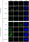
Overexpression of TRF2ΔB induces TIFs in normal but not WS fibroblasts. 53BP1 foci and colocalization with the telomeric protein TRF1 in normal and WS fibroblasts 1 day after transduction of lentiviruses expressing the indicated proteins were detected with antibodies against 53BP1 (green) and TRF1 (red). Nuclei were visualized by DAPI staining. Arrows point at regions of 53BP1 and TRF1 colocalization. For the quantitation of 53BP1 foci and 53BP1 and TRF1 colocalization, see Fig. S1 in the supplemental material.
FIG. 4.
FIG. 4.
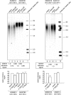
TRF2ΔB induces telomere shortening in normal but not WS fibroblasts. Normal and WS fibroblasts expressing Flag-TRF2ΔB (lanes 2, 5, 9, and 11) and Flag-TRF2 (lanes 3 and 6), along with normal and WS fibroblasts transduced with control viruses (lanes 1, 4, 8, and 10), were harvested 8 days after lentivirus transduction. Equal amounts of genomic DNA digested with HinfI and RsaI were separated by electrophoresis on a 0.8% agarose gel and analyzed by Southern blotting with a radiolabeled (TTAGGG)3 probe. The molecular mass standards shown on right side were generated by digestion of lambda DNA with restriction endonuclease HindIII. Southern blot analyses were performed on three independent samples of normal and WS cells transduced with lentiviruses expressing the indicated proteins. The telomeric signal was normalized to the H1.1 gene probe for all lanes (see Fig. S2 in the supplemental material), and the normalized values ± standard deviations, expressed as the telomeric signal relative to the vector control for each cell line, from three independent experiments (n = 3) are shown below the blots.
FIG. 5.
FIG. 5.
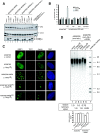
TRF2ΔB-induced cell senescence, TIFs, and telomere shortening are reconstituted in WS fibroblasts genetically complemented with wild-type WRN but not enzymatically deficient WRN variants. (A) Expression of wild-type and mutant forms of WRN in WS cells. WS cells were infected with lentiviruses for the expression wild-type, helicase-deficient, exonuclease-deficient, and helicase- and exonuclease-deficient forms of WRN and cultured for 2 weeks. The parental and genetically complemented cells lines were then transduced with a control virus or a virus for the expression of Flag-TRF2ΔB or Flag-TRF2. Analysis of protein expression was performed by preparation of nuclear extracts, followed by Western blotting with anti-WRN (top panel), antitubulin (middle panel), and anti-Flag (bottom panel) antibodies. (B) Detection of SA-βgal activity. Telomerase-positive WS fibroblasts transduced with the indicated lentiviruses were cultured for 8 days, fixed, and stained for SA-βgal. Five hundred cells of each line were analyzed in duplicate plates. Each bar represents the mean ± the standard deviation of three independent experiments (n = 3) carried out in duplicate. WT, wild type. (C) Detection of 53BP1 and TRF1 in WS fibroblasts consecutively transduced with lentiviruses expressing the indicated proteins with antibodies against 53BP1 (green) and TRF1 (red) 1 day after the second transduction. For the quantitation of 53BP1 foci and 53BP1 and TRF1 colocalization, see Fig. S1 in the supplemental material. (D) The parental and genetically complemented cell lines were transduced with control lentivirus (lanes 1 and 4) and lentiviruses for the expression of Flag-TRF2ΔB (lanes 2 and 5) or Flag-TRF2 (lanes 3 and 6). Cells were harvested 8 days after lentivirus transduction, and genomic DNA was isolated and digested with HinfI and RsaI. Equal amounts (2 μg) of digested genomic DNA were separated by electrophoresis on a 0.8% agarose gel, followed by Southern blot analysis with a radiolabeled (TTAGGG)3 probe. Southern blot analyses were performed on three independent samples of WS cells transduced with lentiviruses expressing the indicated proteins. The telomeric signal was normalized to the H1.1 gene probe for all lanes (see Fig. S2 in the supplemental material), and the normalized values ± standard deviations, expressed as the telomeric signal relative to the vector control for each cell line, from three independent experiments (n = 3) are shown below the blots.
FIG. 6.
FIG. 6.
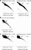
Telomeric circles are present in telomerase-positive WS fibroblasts in the absence of TRF2ΔB. DNA isolated from normal (A) and WS (C) fibroblasts transduced with lentiviruses expressing the indicated proteins was digested with HinfI and RsaI, separated by size and shape, blotted, and probed with a telomeric (CCCTAA) repeat probe. Arrows indicate arcs of telomeric DNA circles. Circularized λ × HindIII DNA fragments were used as molecular size markers (the 23- and 4.4-kb fragments have one cos end and do not circularize). Samples shown in panels A and C were run and processed in parallel under the same hybridization and washing conditions. (B) DNA isolated from ALT fibroblasts was separated by 2DGE and probed with a telomeric (CCCTAA)4 probe. The data shown are representative of at least three independent experiments. The approximate level of telomeric circles (expressed as a percentage of the total telomeric DNA) present in each sample was estimated (see Fig. S4 in the supplemental material) and is shown in the upper right corner of each panel. The samples shown in each panel were blotted, hybridized, washed, and analyzed simultaneously.
FIG. 7.
FIG. 7.
Expression of wild-type WRN but not enzymatically deficient WRN variants in WS fibroblasts leads to a reduction in telomeric circles, which are reformed upon the overexpression of TRF2ΔB. (A) DNA isolated from WS fibroblasts transduced with a vector control lentivirus or a lentivirus expressing WRN was digested with HinfI and RsaI, separated by 2DGE, blotted, and probed with a telomeric (CCCTAA)4 probe. (B) DNA isolated from WS fibroblasts transduced with a lentivirus expressing a WRN variant lacking either exonuclease or helicase activity was digested with HinfI and RsaI, separated by 2DGE, blotted, and probed with a telomeric (CCCTAA)4 probe. Arrows show arcs of telomeric DNA circles. (C) DNA isolated from WRN-complemented WS fibroblasts was transduced with a control lentivirus or a lentivirus expressing TRF2ΔB, digested with HinfI and RsaI, separated by 2DGE, and probed with a telomeric (CCCTAA)4 probe. The samples shown in each panel were run and processed in parallel under the same hybridization and washing conditions. The approximate level of telomeric circles present in each sample (expressed as a percentage of the total telomeric DNA) was estimated (see Fig. S4 in the supplemental material) and is shown in the upper right corner of each panel. The samples shown in each panel were blotted, hybridized, washed, and analyzed simultaneously.
FIG. 8.
FIG. 8.
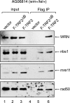
The amino-terminal domain of TRF2 is required for binding to WRN. (A) Nuclear extracts were prepared from normal fibroblasts transduced with a lentivirus for the expression of Flag-TRF2 or Flag-TRF2ΔB and incubated with anti-Flag resin. After extensive washes, the immunoprecipitated proteins were eluted from the beads by treatment with high-salt buffer, separated by 8% SDS-polyacrylamide gel electrophoresis, and analyzed by Western blotting with antibodies against WRN, Nbs1, Mre11, and Rad50 (lanes 4 to 6). Lanes 1 to 3 represent 10% of the nuclear extracts used in the immunoprecipitations (IP) of extracts prepared from control cells (lane 1), cells expressing Flag-TRF2ΔB (lane 2), and cells expressing Flag-TRF2 (lane 3) fibroblasts. Arrows show the migration of relevant proteins.
FIG. 9.
FIG. 9.
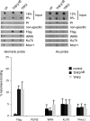
Telomeric association of WRN in fibroblasts is influenced by overexpression of Flag-TRF2 or Flag-TRF2ΔB. Extracts prepared from formaldehyde-treated normal and WS cells expressing Flag-TRF2, Flag-TRF2ΔB, and the vector control were subjected to ChIPs with the indicated antibodies. Coprecipitated DNA were released from the immune complex and analyzed by dot blot hybridization with a radiolabeled (TTAGGG)5 probe. The top panel shows representative dot blots of CHIP assays of cell lines transduced with control (ctr) and TRF2ΔB- and TRF2-specific lentiviruses. The indicated amounts of total input DNA and ChIP DNA were hybridized to telomere-specific and Alu-specific probes. The bottom panel shows results that were visualized by PhosphorImager and quantitated with ImageQuant software (Molecular Dynamics) and are expressed in relative binding units. PI and NS represent control immunoprecipitation reactions with preimmune serum (PI) and a nonspecific (NS) antibody against glutathione S-transferase. Data represent the mean and standard deviation from four independent ChIP assays (n = 4).

Similar articles

Cited by

References

    1. Bilaud, T., C. Brun, K. Ancelin, C. E. Koering, T. Laroche, and E. Gilson. 1997. Telomeric localization of TRF2, a novel human telobox protein. Nat. Genet. 17236-239. - PubMed
    1. Brewer, B. J., and W. L. Fangman. 1987. The localization of replication origins on ARS plasmids in S. cerevisiae. Cell 51463-471. - PubMed
    1. Broccoli, D., A. Smogorzewska, L. Chong, and T. de Lange. 1997. Human telomeres contain two distinct Myb-related proteins, TRF1 and TRF2. Nat. Genet. 17231-235. - PubMed
    1. Chong, L., B. van Steensel, D. Broccoli, H. Erdjument-Bromage, J. Hanish, P. Tempst, and T. de Lange. 1995. A human telomeric protein. Science 2701663-1667. - PubMed
    1. Cohen, S., and S. Lavi. 1996. Induction of circles of heterogeneous sizes in carcinogen-treated cells: two-dimensional gel analysis of circular DNA molecules. Mol. Cell. Biol. 162002-2014. - PMC - PubMed

Publication types

MeSH terms