. 2008 Apr;76(4):1639-48.
doi: 10.1128/IAI.01621-07. Epub 2008 Jan 22.

Genomic analysis reveals Mycoplasma pneumoniae repetitive element 1-mediated recombination in a clinical isolate

Affiliations

Genomic analysis reveals Mycoplasma pneumoniae repetitive element 1-mediated recombination in a clinical isolate

Oxana Musatovova et al. Infect Immun. 2008 Apr.

Abstract

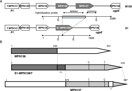
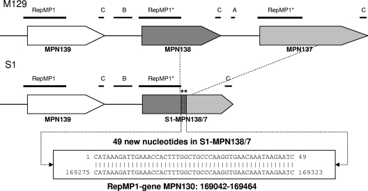
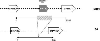
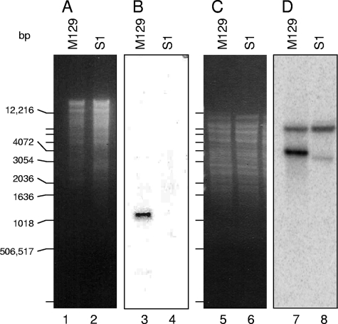
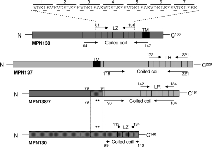
Mycoplasmas are cell wall-less bacteria that evolved by drastic reduction of the genome size. Complete genome analysis of Mycoplasma pneumoniae revealed the presence of numerous copies of four distinct large M. pneumoniae repetitive elements (RepMPs). One copy each of RepMP2/3, RepMP4, and RepMP5 are localized within the P1 operon (MPN140 to MPN142 loci), and their involvement in sequence variation in adhesin P1 and adherence-related protein B/C has been documented. Here we analyzed a clinical strain of M. pneumoniae designated S1 isolated from a 1993 outbreak of respiratory infections in San Antonio, TX. Based on the type of RepMPs within the P1 operon, we classified clinical isolate S1 as type 2 with unique minor sequence variations. Hybridization with oligonucleotide arrays revealed sequence divergence in two previously unsuspected hypothetical genes (MPN137 and MPN138 loci). Closer inspection of this region revealed that the MPN137 and MPN138 loci harbored previously unrecognized unique RepMP1 sequences found only in M. pneumoniae. PCR and sequence analyses revealed a recombination event involving three RepMP1-containing genes that resulted in fusion of MPN137 and MPN138 reading frames and loss of all but a short fragment of another RepMP1-containing locus, MPN130. The multiple copies of unique RepMP1 elements spread throughout the chromosome could allow vast numbers of sequence variations in clinical strains. Comparisons of amino acid sequences showed the presence of leucine zipper motifs in MPN130 and MPN138 proteins in reference strain M129 and the absence of these motifs in the fused protein of S1. The presence of tandem leucine and other repeats points to possible regulatory functions of proteins encoded by RepMP1-containing genes.


Figures

FIG. 1.
FIG. 1.
(A) Comparison of MPN136-MPN141 regions in M. pneumoniae strains M129 and S1. The reference strain M129 region contains the MPN139, MPN138, and MPN137 loci. The three-gene operon containing the adhesin P1 gene (MPN141) is located upstream of the MPN139 locus, and the gene encoding a putative permease (ugpE or MPN136) is located immediately downstream of the MPN137 locus. The MPN138/7 fused reading frame (striped bar, deleted sequence) present in clinical strain S1 is surrounded by conserved sequences. The exact positions of PCR-amplified regions and their sizes are indicated. EcoRI restriction sites (E) are indicated together with the positions of two hybridization probes used in Southern analysis (designated “deleted” and “common”). (B) DNA sequence homologies between S1-MPN138/7 and M129-MPN137 and M129-MPN138 reading frames. The fused S1-MPN138/7 region contains the 5′ end of MPN138 and the 3′ end of MPN137. Two nucleotide transitions are indicated, as are 21 nucleotides missing in S1-MPN138/7 (one asterisk). The position of 49 novel nucleotides is indicated by two asterisks.
FIG. 2.
FIG. 2.
Comparison of RepMP1 elements in the MPN139, MPN138, and MPN137 regions in strains M129 and S1. For M129, the positions of all three RepMP1 core sequences are indicated (asterisks indicate repeats identified in this study). Short repeats (A, B, and C) accompanying RepMP1 core elements are also indicated. For S1, only two copies of the RepMP1 core repeat element were found. The deleted region is indicated by dotted lines. The MPN138/7 fused reading frame contains a proximal region of MPN138, a distal region of MPN137, and a central region consisting of 49 nucleotides previously not found in either locus (two asterisks). BLAST analysis revealed a perfect match with the RepMP1 core element of the MPN130 locus.
FIG. 3.
FIG. 3.
Characterization of PCR-amplified MPN129-MPN131 regions in M. pneumoniae strains M129 and S1. The position of the RepMP1 core element within M129-MPN130 is indicated. In clinical strain S1, the MPN130 locus is deleted (the 680-bp missing region is indicated by a striped bar). The exact positions of PCR-amplified regions and their sizes are indicated.
FIG. 4.
FIG. 4.
Southern analysis of strains M129 and S1. Chromosomal DNA isolated from M129 (lanes 1, 3, 5, and 7) and S1 (lanes 2, 4, 6, and 8) was digested to completion with EcoRI and HindIII, and the fragments generated were separated on 1% agarose gels (A and C). After the restricted DNA was blotted on membranes, Southern hybridization was performed using the deleted portion of the MPN138/7 region as a probe (B), as well as a 32P-labeled MPN130 oligomer (D). A 1-kb DNA ladder was used to estimate the sizes of fragments.
FIG. 5.
FIG. 5.
Predicted secondary structure and leucine-containing tandem repeats in modified proteins encoded by genes containing RepMP1. Regions of coiled coils within the MPN138, MPN137, MPN138/7, and MPN130 proteins are indicated. The positions of LZs in the MPN138 and MPN130 proteins are also indicated. In the LZ motif in the MPN138 protein the positions of leucine and hydrophobic residues (both underlined) are indicated. The locations of LRs in the MPN138 and MPN138/7 proteins are indicated. The positions of transmembrane domains (TM) predicted by dense alignment surface analysis in the MPN137 and MPN138 proteins are also shown. As shown, only the indicated 16-amino-acid region of the MPN130 protein (two asterisks) is retained in S1.

References

    1. Barr, F. G., N. Galili, J. Holick, J. A. Biegel, G. Rovera, and B. S. Emanuel. 1993. Rearrangement of the PAX3 paired box gene in the paediatric solid tumour alveolar rhabdomyosarcoma. Nat. Genet. 3113-117. - PubMed
    1. Baseman, J. B., and J. G. Tully. 1997. Mycoplasmas: sophisticated, reemerging, and burdened by their notoriety. Emerg. Infect. Dis. 321-32. - PMC - PubMed
    1. Buckland, R., and F. Wild. 1989. Leucine zipper motif extends. Nature 338547. - PubMed
    1. Cousin, A., B. de Barbeyrac, A. Charron, H. Renadin, and C. Bebear. 1994. Analysis of RFLPs of amplified cytadhesin P1 gene for epidemiological study of Mycoplasma pneumoniae. IOM Lett. 3494-495.
    1. Cousin-Allery, A., A. Charron, B. de Barbeyrac, G. Fremy, J. Skov Jensen, H. Renaudin, and C. Bebear. 2000. Molecular typing of Mycoplasma pneumoniae strains by PCR-based methods and pulsed-field gel electrophoresis. Application to French and Danish isolates. Epidemiol. Infect. 124103-111. - PMC - PubMed

Publication types

MeSH terms

Substances

Associated data

LinkOut - more resources