. 2008 Mar;76(3):916-26.
doi: 10.1128/IAI.01011-07. Epub 2008 Jan 22.

Tumor necrosis factor blockade in chronic murine tuberculosis enhances granulomatous inflammation and disorganizes granulomas in the lungs

Affiliations

Tumor necrosis factor blockade in chronic murine tuberculosis enhances granulomatous inflammation and disorganizes granulomas in the lungs

Soumya D Chakravarty et al. Infect Immun. 2008 Mar.

Abstract

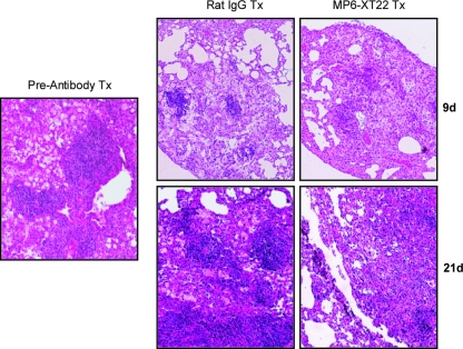
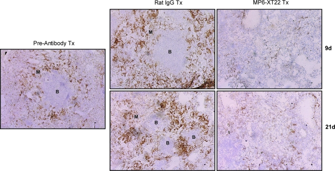
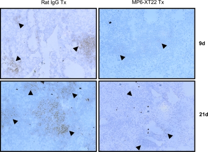
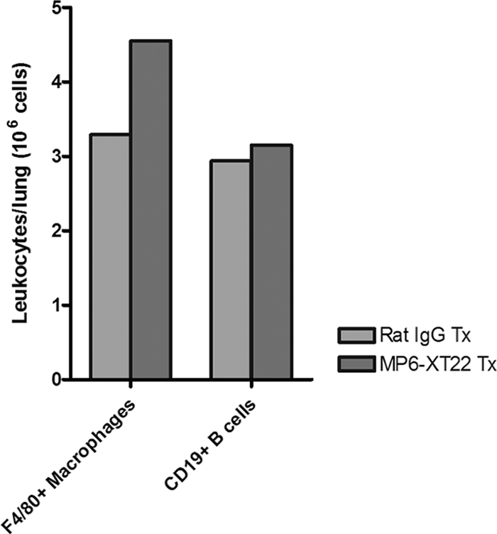
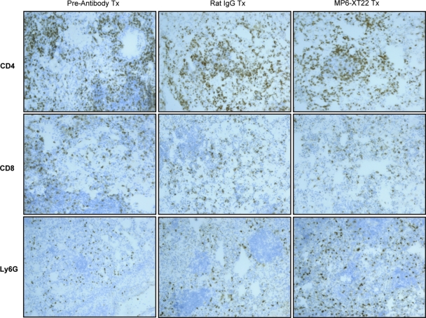
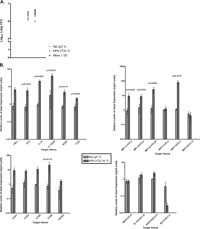
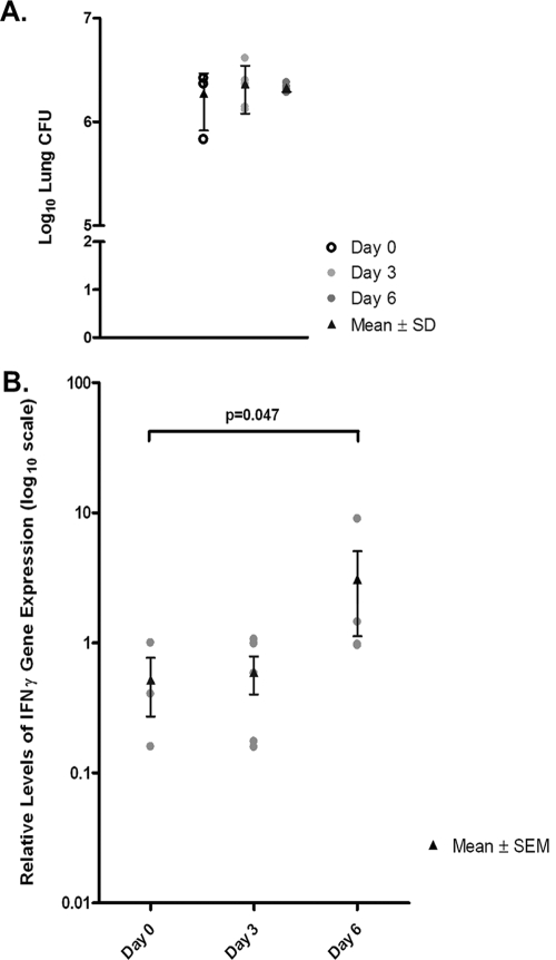
Tumor necrosis factor (TNF) is a prototypic proinflammatory cytokine that contributes significantly to the development of immunopathology in various disease states. A complication of TNF blockade therapy, which is used increasingly for the treatment of chronic inflammatory diseases, is the reactivation of latent tuberculosis. This study used a low-dose aerogenic model of murine tuberculosis to analyze the effect of TNF neutralization on disease progression in mice with chronic tuberculous infections. Histological, immunohistochemical, and flow cytometric analyses of Mycobacterium tuberculosis-infected lung tissues revealed that the neutralization of TNF results in marked disorganization of the tuberculous granuloma, as demonstrated by the dissolution of the previously described B-cell-macrophage unit in granulomatous tissues as well as by increased inflammatory cell infiltration. Quantitative gene expression studies using laser capture microdissected granulomatous lung tissues revealed that TNF blockade in mice chronically infected with M. tuberculosis leads to the enhanced expression of specific proinflammatory molecules. Collectively, these studies have provided evidence suggesting that in the chronic phase of M. tuberculosis infection, TNF is essential for maintaining the structure of the tuberculous granuloma and may regulate the granulomatous response by exerting an anti-inflammatory effect through modulation of the expression of proinflammatory mediators.


Figures

FIG. 1.
FIG. 1.
Lung histopathology of MP6-XT22 and control rat IgG-treated C57BL/6 mice chronically infected with M. tuberculosis. Lungs were harvested from mice at 6 months postinfection prior to the administration of the TNF-neutralizing MP6-XT22 and then at 9 days (9d) and 21 days (21d) posttreatment (Tx); lung samples were formalin fixed, paraffin embedded, sectioned, and H&E stained. Total magnification, ×100. Samples are representative of sections from three or four mice per treatment group per time point (three sections per mouse). The experiment was repeated twice with similar results.
FIG. 2.
FIG. 2.
Immunohistochemical analysis of the effects of TNF neutralization on the B-cell aggregates-macrophage units in the lungs of C57BL/6 mice persistently infected with M. tuberculosis. Lungs were obtained from mice at 6 months postinfection prior to the administration of MP6-XT22, and subsequently at 9 days (9d) and 21 days (21d) posttreatment (Tx), optimal cutting temperature embedded, cryosectioned, and stained for macrophages (F4/80+) to identify the B-cell aggregate-macrophage subunit. Control mice received rat IgG treatment. Virtually all lymphoid aggregates in the lungs of mice with chronic tuberculosis are B-cell nodules (38). “B” denotes aggregates of B lymphocytes; macrophages are designated by “M.” Total magnification, ×100. Samples are representative of sections from three or four mice per treatment group per time point (three sections per mouse). The study was repeated once with similar results.
FIG. 3.
FIG. 3.
Disaggregation of CD19+ B-cell aggregates (brown staining) in the lungs of mice with chronic M. tuberculosis upon TNF neutralization by the MP6-XT22-neutralizing antibody at days 9 and 21 after initiation of treatment. Robust CD19+ B-cell nodules (arrowheads) are seen in the lungs of mice treated with control rat IgG. The lungs of mice treated with MP6-XT22 for 9 and 21 days demonstrate the disaggregation of CD19+ B-cell aggregates but still display remnants of B cells (arrowheads). Photomicrographs are representative and are at ×200 total magnification of 6-μm frozen sections immunoreacted to anti-CD19 antibodies.
FIG. 4.
FIG. 4.
Quantification of F4/80+ macrophages and CD19+ B cells among CD45+ leukocytes obtained from lungs of MP6-XT22- and control rat IgG-treated mice with chronic tuberculosis through flow cytometry. Dead cells were identified and excluded from analysis by means of staining with the Live/Dead reduced biohazard cell viability kit number 2 (green staining) or number 4 (blue staining). Positively enriched live CD45+ cells were obtained and subjected to staining using antibodies for cell surface markers of interest. Absolute numbers of CD19+ B cells and F4/80+ macrophages at 9 days (9d) posttreatment are shown. Proportions of CD45+ cells analyzed were comparable between the two groups. Bars represent data from lung cells pooled from four or five mice. The experiments were repeated once with similar results.
FIG. 5.
FIG. 5.
Immunohistochemical staining of cryosections for T-lymphocyte subsets and neutrophils in M. tuberculosis-infected C57BL/6 mice undergoing TNF blockade therapy. Lungs were obtained from mice at 6 months postinfection prior to the administration of MP6-XT22 and subsequently at 9 days posttreatment (Tx) (the control group received rat IgG). Lungs were sectioned and stained for CD4+ and CD8+ T cells as well as neutrophils (Ly6G+). Total magnification, ×100. Samples are representative of sections from three or four mice per treatment group per time point (three sections per mouse). The experiment was repeated once with similar results.
FIG. 6.
FIG. 6.
Host gene expression analysis of LCM-procured granulomatous lung tissues from control rat IgG-treated and MP6-XT22-treated mice using real-time PCR. (A) Bacillary load in the lungs of the control rat IgG-treated and MP6-XT22-treated C57BL/6 mice infected with M. tuberculosis. Mice were aerogenically infected with a relatively low inoculum of the virulent Erdman strain of M. tuberculosis (∼150 CFU). Following 6 months postinfection, the mice were sacrificed 6 days after initiation of rat IgG or MP6-XT22 treatment. Lungs were homogenized and plated on 7H10 plates, and colonies were enumerated after 21 days. Data depicted represent the mean CFU obtained from each treatment group (the group treated with control IgG contained three mice, and the group treated with MP6-XT22 contained seven mice), and error bars represent standard deviations. (B) RNA prepared from granulomatous tissues in the lungs of mice at 6 days posttreatment (with control rat IgG or MP6-XT22) was used for cDNA synthesis and subjected to real-time PCR for genes of interest. Data presented represent the mean of results obtained from each treatment group (as described for panel A), and error bars represent standard errors of the means. Genes of interest shown with no P value displayed indicate that the differences observed between the two treatment groups were not statistically significant.
FIG. 7.
FIG. 7.
Gene expression profiling of LCM-procured granulomatous lung tissue from MP6-XT22-treated or untreated mice through real-time PCR. (A) Lung bacterial burdens in MP6-XT22-treated C57BL/6 mice infected with M. tuberculosis. Mice were aerogenically infected with a relatively low inoculum of the virulent Erdman strain of M. tuberculosis (∼120 CFU). At 6 months postinfection, the mice were sacrificed at day 0 (untreated) and at days 3 and 6 after the administration of MP6-XT22. Lungs were homogenized and plated on 7H10 plates, and colonies were enumerated after 21 days. Data presented represent the mean CFU obtained from each time point (three for day 0, five for day 3, and four for day 6), and error bars represent standard deviations. The lung bacterial loads of mice in all experimental groups were comparable. (B) RNA was prepared from LCM-procured granulomatous tissues in the lungs of mice prior to the administration of MP6-XT22 and at 3 and 6 days posttreatment. The RNA was used for cDNA synthesis and subjected to real-time PCR for the IFN-γ gene. Data presented represent the means of results obtained from each time point group (as described for panel A), and error bars represent standard errors of the means. The difference observed in IFN-γ gene expression levels between time points for the day 0 group and the day 6 group was statistically significant (P = 0.047).

References

    1. Algood, H. M., J. Chan, and J. L. Flynn. 2003. Chemokines and tuberculosis. Cytokine Growth Factor Rev. 14467-477. - PubMed
    1. Algood, H. M., P. L. Lin, and J. L. Flynn. 2005. Tumor necrosis factor and chemokine interactions in the formation and maintenance of granulomas in tuberculosis. Clin. Infect. Dis. 41(Suppl. 3)S189-S193. - PubMed
    1. Algood, H. M., P. L. Lin, D. Yankura, A. Jones, J. Chan, and J. L. Flynn. 2004. TNF influences chemokine expression of macrophages in vitro and that of CD11b+ cells in vivo during Mycobacterium tuberculosis infection. J. Immunol. 1726846-6857. - PubMed
    1. Aloisi, F., and R. Pujol-Borrell. 2006. Lymphoid neogenesis in chronic inflammatory diseases. Nat. Rev. Immunol. 6205-217. - PubMed
    1. Bogdan, C., Y. Vodovotz, and C. Nathan. 1991. Macrophage deactivation by interleukin 10. J. Exp. Med. 1741549-1555. - PMC - PubMed

Publication types

MeSH terms