Review
. 2008 Jan 15;7(2):127-34.
doi: 10.4161/cc.7.2.5237. Epub 2007 Oct 26.

Ras nanoclusters: combining digital and analog signaling

Affiliations
Review

Ras nanoclusters: combining digital and analog signaling

Angus Harding et al. Cell Cycle. .

Abstract

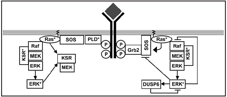
Cellular signaling pathways respond to external inputs to drive pivotal cellular decisions. Far from being mere data relay systems, signaling cascades form complex interacting networks with multiple layers of feedback and feed-forward control loops regulated in both space and time. While it may be intuitively obvious that this complexity allows cells to assess and respond appropriately to a myriad of external cues, untangling the wires to understand precisely how complex networks function as control and computational systems presents a daunting challenge to theoretical and experimental biologists alike. In this review we have focused on activation of the canonical MAP kinase cascade by receptor tyrosine kinases (RTKs) in order to examine some of the fundamental design principles used to build biological circuits and control systems. In particular, we explore how cells can reconfigure signaling cascades to generate distinct biological outputs by utilizing the unique spatial constraints available in biological membranes.


Figures

Figure 1
Figure 1
Multiple regulatory loops exist within the EGF signaling cascade. After the EGF receptor is activated by ligand two exchange factors generate active Ras, the classical Grb2-SOS complex and the recently discovered PLD mediated SOS recruitment. SOS activates Ras, which in turn recruits and activates the MAP kinase module. Multiple regulatory loops exist with the cascade, including a positive feedback loop from active Ras to SOS, a positive feed-forward loop from Ras to KSR, a positive feedback loop from ERK to KSR, a negative feedback loop from ERK to Raf, and a negative feedback loop from ERK back onto itself via the phosphatase DUSP6. The presence of so many regulatory loops provides the pathway with a variety of control systems, which may be used to create different types of output from the Ras pathway in vivo.
Figure 2
Figure 2
One pathway, many outputs. The MAP kinase cascade can produce multiple outputs, including: (A) Graded. Here the system displays a hyperbolic curve, at low-level stimulus the output increases linearly with the input. (B) Switchlike or ultrasensitive. These systems exhibit sigmoidal input-output curves where the first increments produce little response, then when the system does respond it rapidly reaches maximum output. The activation of the MAP kinase module in vitro is switch-like due to the distributive phosphorylation of ERK. (C) Bistable or Digital. These systems represent the extreme conclusion of switch-like kinetics, in which the input-output curve is also sigmoidal and in ideal systems the transition from the off to on state is instantaneous (i.e. the slope of the curve between the off state and the on state is infinite). Although this ideal is never reached in biology, many systems have a sufficiently abrupt transition such that they function as true biological switches to drive discrete cell fate decisions.,,,-
Figure 3
Figure 3
Cytosolic activation of the MAP kinase pathway is suppressed in vivo. Constitutively active human C-Raf mutants (C-Raf with all four activating phosphorylation sites mutated to acidic residues [S338D/Y341D/T491E/ S494D = Raf*] a generous gift from Dr Kun-Liang Guan) were engineered to have different cellular distributions in vivo. Raf* exists in a dynamic equilibrium between the plasma membrane and cytosol due to its membrane-targeting motifs. Raf*ΔM is exclusively cytosolic due to point mutations within each Raf membrane-targeting motifs that abrogates motif-substrate binding (R89L/C165S,C168S/R398A,K399A,R401A).- This mutant was targeted constitutively to the plasma membrane by addition of the C-terminal membrane-targeting motif of K-Ras to generate Raf*ΔM-tK. (A) The schematic diagram shows the cellular distribution expected for each of the mutant proteins. (B) HEK-293 cells transiently transfected and expressing the mutant Raf proteins were harvested, separated into crude membrane and cytosol fractions, and each fraction immunoblotted for C-Raf and ERK2 (=input control). Non-transfected and empty vector transfections are also shown as control. Each of the mutant Raf proteins displays the expected cellular distribution. (C) HEK-293 cells transiently transfected with the mutant Raf proteins were serum starved overnight, harvested and whole cell lyates immunoblotted for C-Raf, ppERK and ERK2. Strikingly Raf*ΔM, which does not have access to the plasma membrane, was a poor activator of the MAP kinase pathway in vivo. We conclude from these results and others that activation of the MAP kinase pathway by Raf is suppressed within the cytosol.
Figure 4
Figure 4
EGF signal transmission is digitised across the plasma membrane. (A) Ras nanoclusters have an avarage radius of 6–12 nanometers and short lifetimes (~4 s).- During their brief existence, Ras nanoclusters generate maximal ppERK output, functioning as switches (termed nanoswitches to capture their size and digital signal output). (B) EGF is the analog input present in the extracellular matrix (ECM). Activated EGF receptors trigger the formation of Ras nanoswitches at the plasma membrane in direct proportion to the external EGF input through an unknown mechanism. The plasma membrane thus digitizes analog EGF inputs using individual Ras nanoswitches as discrete quanta, functioning as a linear analog-to-digital converter (ADC). As the output of each nanocluster is dumped into the cytoplasm, the digital pulses from individual nanoclusters are summed to give a final analog ppERK output. In this way the cytoplasm reverses the digital conversion that occurred at the plasma membrane and functions as a digital-to-analog converter (DAC). This biological circuit generates the high fidelity signal transmission across the plasma membrane that occurs in vivo.
Figure 5
Figure 5
Fast sampling rates and large sampling precision generates high fidelity signal transmission. The fidelity of signal transmission in a linear ADC is controlled by two variables: the sampling rate, which determines how many samples are taken over time; and the sampling precision, which determines how many quantization levels are between zero and maximum. Figure 5 shows how increasing both these variables increases signal fidelity during signal transmission. The red line represents the analog input signal that changes in amplitude over time. In the upper left panel, both the sampling rate and the sampling precision of the ADC are low (5 and 4 respectively). When the DAC recreates the analog input curve from these numbers it generates the blue line shown in the upper right panel. It is clear from this graph that the output curve is a poor representation on the analog input and much data has been lost during signal transmission. If both the sampling rate and sampling precision of the ADC is increased (bottom panels), then the output curve generated from the DAC more precisely matches the input, generating an equivalent improvement in signal fidelity.
Figure 6
Figure 6
Ras nanoclusters amplify low MEK activity in vivo. COS cells were transfected with constitutively active Raf proteins targeted to the plasma membrane (Raf*-tK) with either full activity [Raf*-tK], catalytically inactive [Raf*-tK(KD)], intermediate catalytic activity (Raf*-tK(S508N)], or very low catalytic activity (Raf*-tK(S508D)], serum starved for 18 hours then whole cell lysates immunoblotted for C-Raf, ppMEK, MEK1, ppERK and ERK2. Raf*-tK activated both MEK and ERK in vivo, whereas the kinase dead control did not. Raf*-tK(S508N) activated MEK equivalent to the fully active control, confirming that nanoclusters amplify low Raf activity in vivo. Raf*-tK(S508D), the Raf protein with the lowest catalytic activity, poorly activated MEK. Nevertheless, this low MEK activity was sufficient to fully activate ERK in vivo. Thus both in silico and in vivo experiments support the idea that Ras nanoclusters amplify both Raf and MEK activity, functioning as high-gain amplifiers for the MAP kinase module in vivo.
Figure 7
Figure 7
RKIP inhibits cytosolic but not plasma membrane activation of the MAPK module. (A) COS cells transfected with constitutively active Raf-1 (Raf*) or constitutively active Raf targeted to the plasma membrane (Raf*-tK) with or without RKIP were serum starved, fractionated and the membrane and cytosolic fractions immunoblotted for C-Raf. The cytosolic fractions were immunoblotted for ppERK, ERK2 and RKIP. RKIP expression efficiently inhibited ERK activation by Raf*, whereas RKIP did not inhibit ERK activation by membrane targeted Raf*-tK. (B) Identical aliquots of COS cells were co-transfected with 5 μg of either Raf* or Raf*-tK plasmid, and a range of RKIP plasmid concentrations (0–20 μg). After 48 hours cells were harvested and proliferation quantified by counting cells. Figure 6B shows that even at high levels of RKIP expression, cells expressing membrane targeted Raf*-tK continued to proliferate. In contrast, low levels of RKIP expression inhibited proliferation of cells expressing cytosolic Raf* (The RKIP expression vector was a generous gift from Dr. Walter Kolch68).

Similar articles

Cited by

References

    1. Schlessinger J. Cell signaling by receptor tyrosine kinases. Cell. 2000;103:211–25. - PubMed
    1. Pawson T. Regulation and targets of receptor tyrosine kinases. Eur J Cancer. 2002;38:3–10. - PubMed
    1. McKay MM, Morrison DK. Integrating signals from RTKs to ERK/MAPK. Oncogene. 2007;26:3113–21. - PubMed
    1. Hancock JF, Magee AI, Childs JE, Marshall CJ. All ras proteins are polyisoprenylated but only some are palmitoylated. Cell. 1989;57:1167–77. - PubMed
    1. Hancock JF, Paterson H, Marshall CJ. A polybasic domain or palmitoylation is required in addition to the CAAX motif to localize p21ras to the plasma membrane. Cell. 1990;63:133–9. - PubMed

Publication types

MeSH terms

Substances

LinkOut - more resources