

Taxol crystals can masquerade as stabilized microtubules
- PMID: 18213384
- PMCID: PMC2194920
- DOI: 10.1371/journal.pone.0001476
Taxol crystals can masquerade as stabilized microtubules
Abstract
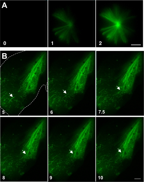
Taxol is a potent anti-mitotic drug used in chemotherapy, angioplastic stents, and cell biology research. By binding and stabilizing microtubules, Taxol inhibits their dynamics, crucial for cell division, motility, and survival. The drug has also been reported to induce formation of asters and bundles composed of stabilized microtubules. Surprisingly, at commonly used concentrations, Taxol forms crystals that rapidly bind fluorescent tubulin subunits, generating structures with an uncanny resemblance to microtubule asters and bundles. Kinetic and topological considerations suggest that tubulin subunits, rather than microtubules, bind the crystals. This sequestration of tubulin from the subunit pool would be expected to shift the equilibrium of free to polymerized tubulin to disfavor assembly. Our results imply that some previously reported Taxol-induced asters or bundles could include or be composed of tubulin-decorated Taxol crystals. Thus, reevaluation of certain morphological, chemical, and physical properties of Taxol-treated microtubules may be necessary. Moreover, our findings suggest a novel mechanism for chemotherapy-induced cytotoxicity in non-dividing cells, with far-reaching medical implications.
Conflict of interest statement
Figures


Similar articles
-
Demonstration of the cell cycle positions of taxol-induced "asters" and "bundles" by sequential measurements of tubulin immunofluorescence, DNA content, and autoradiographic labeling of taxol-sensitive and -resistant cells.J Histochem Cytochem. 1989 Nov;37(11):1659-65. doi: 10.1177/37.11.2572626. J Histochem Cytochem. 1989. PMID: 2572626
-
Sulfo-SMCC Prevents Annealing of Taxol-Stabilized Microtubules In Vitro.PLoS One. 2016 Aug 25;11(8):e0161623. doi: 10.1371/journal.pone.0161623. eCollection 2016. PLoS One. 2016. PMID: 27561096 Free PMC article.
-
Tau induces cooperative Taxol binding to microtubules.Proc Natl Acad Sci U S A. 2004 Aug 31;101(35):12910-5. doi: 10.1073/pnas.0402928101. Epub 2004 Aug 23. Proc Natl Acad Sci U S A. 2004. PMID: 15326286 Free PMC article.
-
Taxol®: The First Microtubule Stabilizing Agent.Int J Mol Sci. 2017 Aug 9;18(8):1733. doi: 10.3390/ijms18081733. Int J Mol Sci. 2017. PMID: 28792473 Free PMC article. Review.
-
Effects of a microtubule stabilizing agent on the response of platelets to vincristine.Blood. 1982 Aug;60(2):474-83. Blood. 1982. PMID: 6124288 Review.
Cited by
-
Mathematical modeling of microtubule dynamics: insights into physiology and disease.Prog Neurobiol. 2010 Dec;92(4):478-83. doi: 10.1016/j.pneurobio.2010.08.003. Epub 2010 Aug 14. Prog Neurobiol. 2010. PMID: 20713128 Free PMC article.
-
Chromosomal instability reducing effect of paclitaxel and lapatinib in mouse embryonic stem cells with chromosomal abnormality.Mol Biol Rep. 2020 Nov;47(11):8605-8614. doi: 10.1007/s11033-020-05903-8. Epub 2020 Oct 15. Mol Biol Rep. 2020. PMID: 33057993
-
Radial alignment of microtubules through tubulin polymerization in an evaporating droplet.PLoS One. 2020 Apr 10;15(4):e0231352. doi: 10.1371/journal.pone.0231352. eCollection 2020. PLoS One. 2020. PMID: 32275729 Free PMC article.
-
Effect of (2)H and (18)O water isotopes in kinesin-1 gliding assay.PeerJ. 2014 Mar 25;2:e284. doi: 10.7717/peerj.284. eCollection 2014. PeerJ. 2014. PMID: 24711961 Free PMC article.
-
Paclitaxel and TRAIL synergize to kill paclitaxel-resistant small cell lung cancer cells through a caspase-independent mechanism mediated through AIF.Anticancer Res. 2011 Oct;31(10):3193-204. Anticancer Res. 2011. PMID: 21965726 Free PMC article.
References
-
- Wani MC, Taylor HL, Wall ME, Coggon P, McPhail AT. Plant antitumor agents. VI. The isolation and structure of taxol, a novel antileukemic and antitumor agent from Taxus brevifolia. J Am Chem Soc. 1971;93:2325–2327. - PubMed
-
- Derry WB, Wilson L, Jordan MA. Substoichiometric binding of taxol suppresses microtubule dynamics. Biochemistry. 1995;34:2203–2211. - PubMed
-
- Nogales E, Whittaker M, Milligan RA, Downing KH. High-resolution model of the microtubule. Cell. 1999;96:79–88. - PubMed
Publication types
MeSH terms
Substances
LinkOut - more resources
Full Text Sources
