

doi: 10.1371/journal.pone.0001477.
Mammalian cells change volume during mitosis
Affiliations
- PMID: 18213385
- PMCID: PMC2198941
- DOI: 10.1371/journal.pone.0001477
Item in Clipboard
Mammalian cells change volume during mitosis
PLoS One.
.
Abstract
Using single cell-imaging methods we have found that the volume of adherent cells grown in culture decreases as the cells rounds when it enters mitosis. A minimal volume is reached at metaphase. Rapid volume recovery initiates before abscission as cells make the transition from metaphase to cytokinesis. These volume changes are simultaneous with the rapid surface area decrease and recovery observed in mitotic cells [1].
Conflict of interest statement
Figures
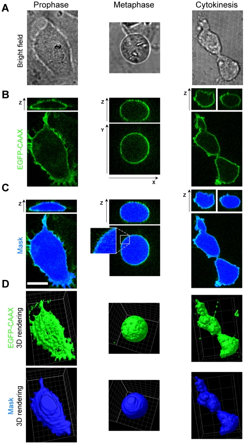
(A) Representative images of the same BSC1 cell visualized by bright field phase contrast illumination during prophase, metaphase or cytokinesis (B) Ortogonal fluorescent views corresponding to the distribution of EGFP-CAAX in the same cell imaged using the confocal spinning head. The stacks along the z-axis correspond to sequential optical sections acquired 0.25 µm apart. The panels show the fluorescence signal along the z-axis (top) along a single confocal plane positioned approximately in the middle of the cell. Bar, 10 µm. (C) The mask (light blue) corresponds to the position of the EGFP-CAAX signal calculated for each confocal plane (see Methods). The outline of the outer boundary determined for any given plane defines the enclosed area for such plane (dark blue). (D) Three-dimensional rendition comparing the distribution of EGFP-CAAX (green) and the calculated cell volume (blue).
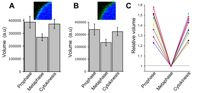
(A) Population analysis (average +/− standard deviation) of cell volume determined in 6 experiments for 10 single cells imaged during prophase, metaphase and last stages of cytokinesis. The most external pixels of the EGFP-CAAX signal were used to generate the mask (inset). The statistical significance of the volume difference between the cell cycle stages was calculated using the t-Student test (p<0.001). (B) Same analysis than in (A), but with the most internal pixels of the EGFP-CAAX signal were used to generate the mask (inset). (C) Individual representation of the volume data presented in (A). To facilitate the comparisons, the volume of each cell during metaphase was normalized to a relative value of 1.
References
-
- Wright LP, Philips MR. Thematic review series: lipid posttranslational modifications. CAAX modification and membrane targeting of Ras. J Lipid Res. 2006;47(5):883–891. - PubMed
-
- Schneider EL, Fowlkes BJ. Measurement of DNA content and cell volume in senescent human fibroblasts utilizing flow multiparameter single cell analysis. Exp Cell Res. 1976;98(2):298–302. - PubMed
Publication types
MeSH terms
Grants and funding
LinkOut - more resources
Full Text Sources
Other Literature Sources
