. 2008 Jan 23;3(1):e1487.
doi: 10.1371/journal.pone.0001487.

Genome-wide and functional annotation of human E3 ubiquitin ligases identifies MULAN, a mitochondrial E3 that regulates the organelle's dynamics and signaling

Affiliations

Genome-wide and functional annotation of human E3 ubiquitin ligases identifies MULAN, a mitochondrial E3 that regulates the organelle's dynamics and signaling

Wei Li et al. PLoS One. .

Abstract

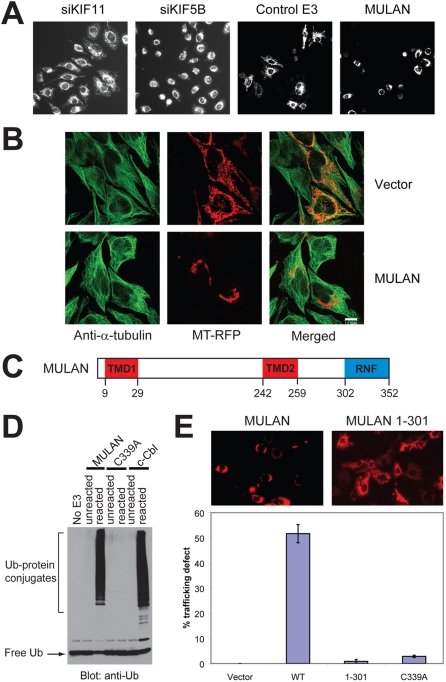
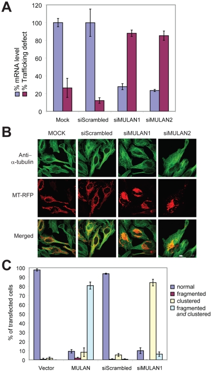
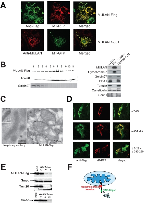
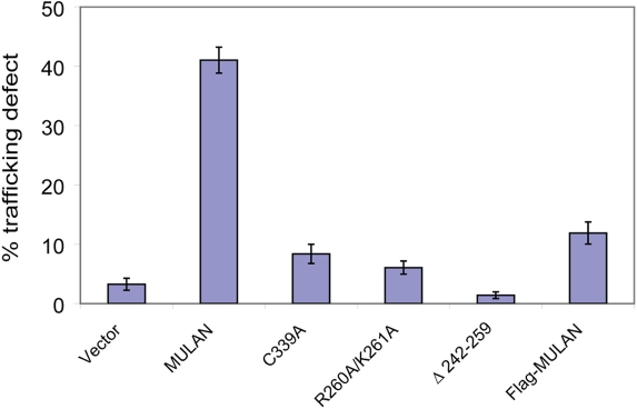
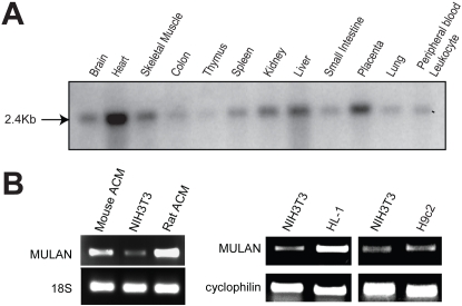
Specificity of protein ubiquitylation is conferred by E3 ubiquitin (Ub) ligases. We have annotated approximately 617 putative E3s and substrate-recognition subunits of E3 complexes encoded in the human genome. The limited knowledge of the function of members of the large E3 superfamily prompted us to generate genome-wide E3 cDNA and RNAi expression libraries designed for functional screening. An imaging-based screen using these libraries to identify E3s that regulate mitochondrial dynamics uncovered MULAN/FLJ12875, a RING finger protein whose ectopic expression and knockdown both interfered with mitochondrial trafficking and morphology. We found that MULAN is a mitochondrial protein - two transmembrane domains mediate its localization to the organelle's outer membrane. MULAN is oriented such that its E3-active, C-terminal RING finger is exposed to the cytosol, where it has access to other components of the Ub system. Both an intact RING finger and the correct subcellular localization were required for regulation of mitochondrial dynamics, suggesting that MULAN's downstream effectors are proteins that are either integral to, or associated with, mitochondria and that become modified with Ub. Interestingly, MULAN had previously been identified as an activator of NF-kappaB, thus providing a link between mitochondrial dynamics and mitochondria-to-nucleus signaling. These findings suggest the existence of a new, Ub-mediated mechanism responsible for integration of mitochondria into the cellular environment.


Conflict of interest statement

Competing Interests: The authors have declared that no competing interests exist.

Figures

Figure 1
Figure 1. Identification of MULAN as a novel regulator of mitochondrial dynamics.
A) Left 2 panels: Mitochondrial perinuclear clustering (a consequence of defective trafficking) results from siRNA-mediated knockdown of KIF5B, but not of KIF11. HeLa cells were transfected with the indicated siRNAs and mitochondria were visualized with MitoTracker Red. Right 2 panels: A cell-based imaging screen of E3 cDNA and shDNA genome-wide collections led to the identification of MULAN, a RING finger protein whose ectopic expression resulted in mitochondrial perinuclear clustering and fragmentation. For the screen, HeLa cells were co-transfected with mitochondrial-targeted RFP (MT-RFP) and with the E3 collections. “Control E3” refers to a random E3 cDNA which did not affect mitochondrial dynamics in the screen. B) Mitochondrial perinuclear clustering in response to MULAN ectopic expression apparently does not result from disruption of the microtubule network. HeLa cells were co-transfected with MT-RFP and either vector control or MULAN wild type cDNA. Cells were immuno-stained with anti-α-tubulin antibody to visualize the microtubule network. Green, tubulin staining; red, mitochondria. C) MULAN's schematic domain structure. Amino acid numbers indicated below. TMD, transmembrane domain. RNF, RING finger. D) MULAN's RNF has in vitro E3 activity. Reactions utilized GST fusions with the MULAN or c-Cbl RNFs as E3s. Activity was dependent on an intact RNF, since it was not detected in reactions using the MULAN C339A RNF mutant. c-Cbl's RNF was used as a positive control. Negative controls were reactions lacking an E3 (No E3) or reactions added to SDS sample buffer at t = 0 (unreacted). E) MULAN's RNF is required for the regulation of mitochondrial dynamics. NIH3T3 cells were transfected with vector, wild type MULAN cDNA or RNF-mutant cDNAs, together with MT-RFP. Cells were fixed at 24 h post-transfection and the fraction of RFP-positive cells exhibiting perinuclear-clustered mitochondria was scored (% trafficking defect). Data represent the average of at least 350 cells per condition, from 3–5 random 20× fields. Results are representative of several experiments.
Figure 2
Figure 2. Knockdown of endogenous MULAN perturbs mitochondrial dynamics.
A) Blue bars: MULAN mRNA knockdown. RNA was extracted from HeLa cells 48 h after transfection with siRNA oligos targeting two different MULAN sequences, or with a negative control siRNA (siScrambled). Knockdown efficiency was determined by quantitative real-time RT-PCR. Results were normalized to the level of the 36B4 mRNA, which was amplified in the same multiplex reaction, as an internal control. Purple bars: MULAN knockdown leads to mitochondrial perinuclear clustering. HeLa cells were transfected with the indicated siRNAs on day 1, followed by MT-RFP on day 2. Cells were fixed on day 3 and the percentage of RFP-positive cells with perinuclear-clustered mitochondria was scored. B) The microtubule network is apparently intact in MULAN-knockdown cells. As in “A”, except that cells were fixed and stained with anti-α-tubulin antibody for analysis (green). C) Both MULAN ectopic expression and endogenous knockdown indicate its role in mitochondrial dynamics. For ectopic expression, HeLa cells were transfected with vector or MULAN wild type cDNA together with MT-RFP. Cells were fixed for analysis 24 h post-transfection. For siRNA-mediated knockdown, cells were transfected with siScrambled or siMULAN1 on day 1 and with MT-RFP on day 2. Cells were fixed on day 3. Mitochondrial fragmentation and clustering were quantitated under 100× field. At least 100 cells were counted per condition and data represent average of three independent transfections.
Figure 3
Figure 3. MULAN is a mitochondrial outer membrane (MOM) protein with a cytosolic-facing C-terminal RING-finger.
A) MULAN colocalizes with MT-RFP/MT-GFP. NIH3T3 cells were transfected with Flag-tagged MULAN or untagged MULAN 1-301, together with MT-RFP (top panels) or MT-GFP (bottom panels), followed by immunostaining with antibody against Flag (top; green) or MULAN (bottom; red). B) Left panel: MULAN-Flag co-sediments with the mitochondrial protein, Tom20, in sucrose gradient. 293 cells transfected with MULAN-Flag were subfractionated by sucrose gradient. MULAN-Flag, the MOM protein Tom20, and Golgin 97 in each fraction were detected by western blot. Right panel: endogenous MULAN co-fractionates with mitochondria in sucrose gradient. 293F cells were Dounce-homogenized and centrifuged at 750×g to pellet nuclei and unbroken cells. The post-nuclear supernatant was centrifuged at 12,500×g to obtain the heavy membrane (HM) and the cytosolic/light membrane (LM) fractions. The HM fraction was subjected to sucrose gradient to further purify mitochondria. Equal protein amounts (40 µg) were fractionated by SDS-PAGE and blotted with anti-MULAN antibody to detect endogenous MULAN. Cytochrome c, Golgin 97, EEA1 and tubulin were used as markers for mitochondria, Golgi, endosomes and cytosol, respectively. Calreticulin and Sec61 were both used as ER markers. C) Immuno-electron microscopy of MULAN. COS7 cells transfected or not with MULAN-Flag were fixed and stained with anti-Flag antibody followed by gold-conjugated secondary antibody for immuno-EM analysis. Left panel: BSA only (no primary antibody) control; inset: mitochondrion from an untransfected cell. Right panel: mitochondria expressing MULAN-Flag. Scale bars are indicated. D) MULAN's predicted transmembrane domains (TMDs) mediate localization to mitochondria. NIH3T3 cells were co-transfected with Flag-tagged MULAN deletion mutants (green) and MT-RFP (red). Deletion of TMD1 (MULAN Δ2-29; top row) or of TMD2 (MULAN Δ242-259; middle row) led to partial mislocalization of MULAN. The combined deletion of both TMDs led to complete mislocalization of MULAN to the cytosol (MULAN Δ2-29+Δ242-259; bottom row). E) MULAN is a mitochondrial outer membrane (MOM) protein. Mitochondria from 293 cells transfected with MULAN-Flag were purified by sucrose gradient. Intact mitochondria were treated with trypsin for the indicated time, in the presence or absence of Triton X-100. The MULAN C-terminus was readily susceptible to trypsin digestion in intact mitochondria, indicating that it sits in the MOM facing the cytosol. Controls are the MOM protein, Tom20, and the intermembrane space protein, Smac. Smac only becomes sensitive to trypsin upon lysis of mitochondria with 0.5% Triton X-100 (lower panel). F) Topological model for MULAN on the MOM, indicating its transmembrane domains (red) and RNF (green). The cytosolic-exposed RNF can have access to the remaining components of the Ub system.
Figure 4
Figure 4. Modulation of mitochondrial dynamics by MULAN requires its proper localization to the MOM.
NIH3T3 cells were transfected with wild type or mitochondrial localization-defective MULAN mutants, together with MT-RFP. C339A is the RING finger-mutant control. Cells were fixed 24 h post-transfection and the percentage of RFP-positive cells with perinuclear-clustered mitochondria was determined.
Figure 5
Figure 5. MULAN is most highly expressed in the human heart.
A) Full-length human MULAN cDNA was used as a probe for hybridization to northern-blot of RNA from multiple human tissues. A single 2.4Kb band was detected for MULAN mRNA, in various tissues. B) RT-PCR of MULAN mRNA showed higher expression in mouse and rat adult primary cardiomyocytes (ACM) and in the mouse HL-1 cardiomyocyte cell line, compared to mouse NIH-3T3 cells and to the rat skeletal muscle-like cell line H9c2. 18S rRNA and Cyclophilin B were used as controls.

References

    1. Pickart CM, Eddins MJ. Ubiquitin: structures, functions, mechanisms. Biochim Biophys Acta. 2004;1695:55–72. - PubMed
    1. Joazeiro CA, Weissman AM. RING finger proteins: mediators of ubiquitin ligase activity. Cell. 2000;102:549–552. - PubMed
    1. Petroski MD, Deshaies RJ. Function and regulation of cullin-RING ubiquitin ligases. Nat Rev Mol Cell Biol. 2005;6:9–20. - PubMed
    1. Koppen M, Langer T. Protein Degradation within Mitochondria: Versatile Activities of AAA Proteases and Other Peptidases. Crit Rev Biochem Mol Biol. 2007;42:221–42. - PubMed
    1. Fisk HA, Yaffe MP. A role for ubiquitination in mitochondrial inheritance in Saccharomyces cerevisiae. J Cell Biol. 1999;145:1199–208. - PMC - PubMed

Publication types

Substances