Comparative Study
. 2008 Feb;237(2):476-84.
doi: 10.1002/dvdy.21429.

Slits and Robos in the developing chicken inner ear

Affiliations
Comparative Study

Slits and Robos in the developing chicken inner ear

Andrea C Battisti et al. Dev Dyn. 2008 Feb.

Abstract

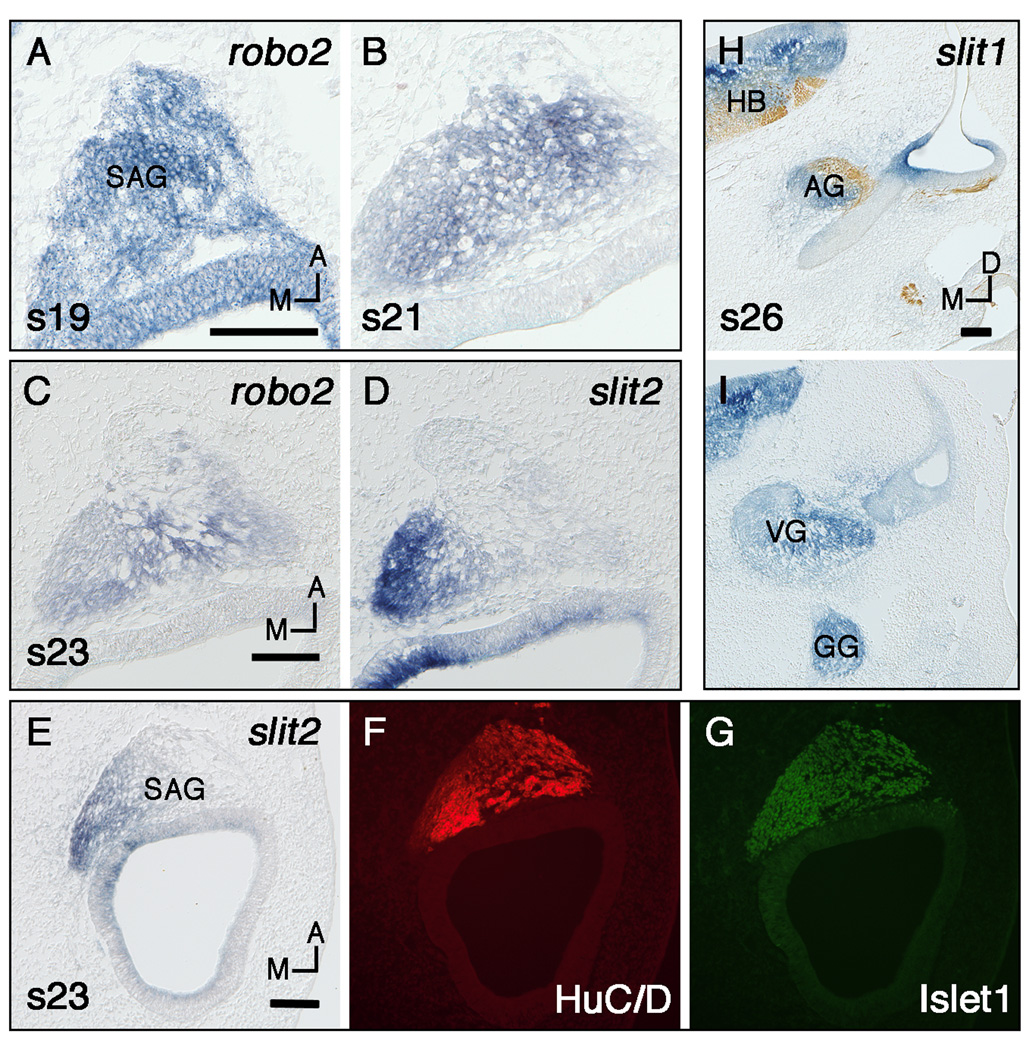
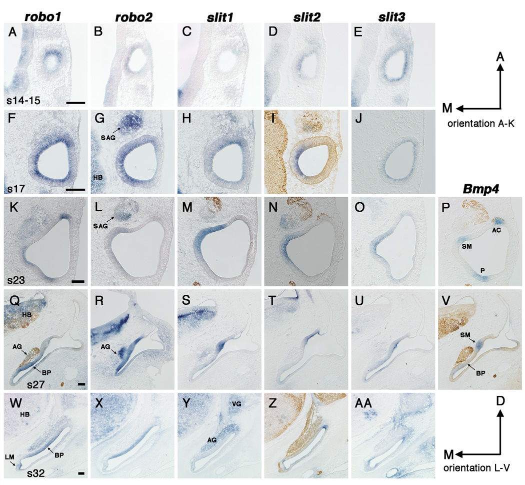
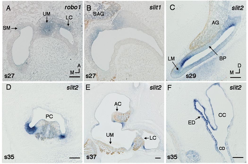
Mechanosensory hair cells in the chick inner ear synapse onto afferent neurons of the statoacoustic ganglion (SAG). During development, these neurons extend a central process to the brain and a peripheral process into one of eight sensory organs. A combination of cues, including chemoattractants and chemorepellents, direct otic axons to their peripheral targets. As a first step in evaluating the role of known axon guidance molecules, Slits and Robos, we examined expression of their transcripts in the chick inner ear from embryonic day 2-11 (Hamburger and Hamilton stages 14-37). Robo2 and slit2 are in migrating neuroblasts and the SAG, while both slits and robos are in the otic epithelium. We speculate that this family of signaling molecules may be involved in repulsion, first of otic neuroblasts and then of otic axons. Later our expression data revealed a potentially novel role for these molecules in maintaining sensory/nonsensory boundaries.


Figures

Fig. 1
Fig. 1
Expression of slits and robos in the statoacoustic ganglion (SAG) determined by in situ hybridization on frozen sections in either the horizontal (A–G) or transverse plane (H–I). Section H is double labeled with a neurofilament antibody. A,B: Robo2 expression in the SAG at stages 19 (A) and 21 (B). C,D: Stage 23 serial sections labeled with robo2 (C) and slit2 (D). E–G: Stage 23 serial sections labeled with slit2 (E) or double labeled with HuC/D and Islet1 antibodies (F,G respectively). H,I: slit1 expression in the auditory (H), vestibular (I), and geniculate ganglia (I) at stage 26. Scale Bars, 100µm. Orientation: A, anterior; D, dorsal; M, medial. Abbreviations: AG, auditory ganglion; GG, geniculate ganglion; HB, hindbrain; VG, vestibular ganglion.
Fig. 2
Fig. 2
Expression of slits and robos in the developing chick inner ear from stage 14 (E2) to stage 32 (E7.5). In situ hybridizations on frozen sections in the horizontal (A–P) or transverse plane (Q–AA). Some sections (I,M,N,P,Q,V,Z) are double labeled with a neurofilament antibody. Prosensory regions are identified by Bmp4 expression (P,V). A–E: Stage 14 (A–C) or stage 15 (D,E), A,C and D,E are serial sections. F–J: Stage 17, F–H are serial sections. K–P: Stage 23, L,M and N,P are serial sections. Q–V: Stage 27, S–V are serial sections. W–AA: Stage 32, W,X,Z and Y,AA are serial sections. Scale Bars, 100µm. Orientation: A, anterior; D, dorsal; M, medial. Abbreviations: AC, anterior crista; AG, auditory ganglion; BP, basilar papilla; E, embryonic day; LM, lagenar macula; HB, hindbrain; P, posterior Bmp4-positive focus; SAG, statoacoustic ganglion; SM, saccular macula; VG, vestibular ganglion.
Fig. 3
Fig. 3
A,B: Robo1 (A) and slit1 (B) in the utricular macula at stage 27 shown by in situ hybridzation on horizonal sections. C–E: Slit2 flanks sensory territories shown in transverse sections. Sections (B,C,E) are double labeled with a neurofilament antibody. Slit2 flanks the auditory basilar papilla (BP) at stage 29 (C). Slit2 flanks the vestibular sensory epithelia, including the posterior crista (PC) at stage 35 (D), and the anterior crista (AC), and the lateral crista (LC) at stage 37 (E). Slit2 is expressed in the endolymphatic duct (ED) at stage 35 (F). Scale Bars, 100µm. Orientation: A, anterior; D, dorsal; M, medial. Abbreviations: CC, common crus; CD, cochlear duct; SM, saccular macula; SAG, statoacoustic ganglion; UM, utricular macula.

References

    1. Bashaw GJ, Goodman CS. Chimeric axon guidance receptors: the cytoplasmic domains of slit and netrin receptors specify attraction versus repulsion. Cell. 1999;97:917–926. - PubMed
    1. Bissonnette JP, Fekete DM. Standard atlas of the gross anatomy of the developing inner ear of the chicken. J Comp Neurol. 1996;368:620–630. - PubMed
    1. Bovolenta P. Morphogen signaling at the vertebrate growth cone: a few cases or a general strategy? J Neurobiol. 2005;64:405–416. - PubMed
    1. Brigande JV, Iten LE, Fekete DM. A fate map of chick otic cup closure reveals lineage boundaries in the dorsal otocyst. Dev Biol. 2000;227:256–270. - PubMed
    1. Brose K, Bland KS, Wang KH, Arnott D, Henzel W, Goodman CS, Tessier-Lavigne M, Kidd T. Slit proteins bind Robo receptors and have an evolutionarily conserved role in repulsive axon guidance. Cell. 1999;96:795–806. - PubMed

Publication types

MeSH terms

Substances