Comparative Study
. 2008 Feb;237(2):504-12.
doi: 10.1002/dvdy.21437.

Genetic targeting of the endoderm with claudin-6CreER

Affiliations
Comparative Study

Genetic targeting of the endoderm with claudin-6CreER

William J Anderson et al. Dev Dyn. 2008 Feb.

Abstract

A full description of the ontogeny of the beta cell would guide efforts to generate beta cells from embryonic stem cells (ESCs). The first step requires an understanding of definitive endoderm: the genes and signals responsible for its specification, proliferation, and patterning. This report describes a global marker of definitive endoderm, Claudin-6 (Cldn6). We report its expression in early development with particular attention to definitive endoderm derivatives. To create a genetic system to drive gene expression throughout the definitive endoderm with both spatial and temporal control, we target the endogenous locus with an inducible Cre recombinase (Cre-ER(T2)) cassette. Cldn6 null mice are viable and fertile with no obvious phenotypic abnormalities. We also report a lineage analysis of the fate of Cldn6-expressing embryonic cells, which is relevant to the development of the pancreas, lung, and liver.


Figures
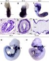
Fig. 1
Fig. 1
Expression of Cldn6 from commencement of gastrulation to the early stages of gut tube organogenesis. A,D: Expression at E6.5 is quite broad throughout the epiblast, with the exception of the primitive streak and visceral endoderm. Similar expression is seen at E7.5 (B,E). C,F: By E8.5, expression begins to be restricted to the endoderm and by E9.5 (G) and E10.5 (H), expression is restricted to the endoderm, otic vesicle (arrowhead), and mesonephros (arrow). A–C are representative whole mount stainings for the sections in D–F), respectively. Embryos in D–F were sectioned in their deciduas. Plane of section is indicated by a line in the corresponding whole mount embryo. For E, posterior is facing up. dec, decidua; ect, ectoderm; mes, mesoderm; end, endoderm; rm, Reichert's membrane; nf, neural fold; he, heart.
Fig. 2
Fig. 2
Epithelial-restricted expression of Cldn6 in the developing lung and pancreas. A–D: Whole mount in situ hybridization shows Cldn6 expression throughout the entire epithelia of the lung from E11.5 to E14.5 [(A) E11.5, (B) E12.5, (C) E13.5, (D) E14.5)]. E,F: Expression of Cldn6 in the E14.5 lung is restricted to the epithelia with no mesenchymal expression. E: Expression of Cldn6 can be seen in the tracheal epithelia, as well as both the proximal and distal airways. F: A higher magnification view of staining from a different lung. Epithelia is outlined by a black dashed line. G: Expression of Cldn6 in the E14.5 pancreas. As with the lung, expression is restricted to the epithelia. Staining in the epithelial lining of the stomach can be seen faintly. H: Cldn6 expression in the E13.5 pancreas. The pancreas is still attached to the stomach. Strong epithelial staining can be seen in the branching epithelia. panc, pancreas; st, stomach.
Fig. 3
Fig. 3
Successful targeting of the Cldn6 locus with the CIHV cassette. A: Southern blot analysis. Upon successful recombination, the entire coding sequence for Cldn6 is removed, generating a null allele. After NcoI digest, the wild type locus generates a band of approximately 12.5 kb, while the targeted allele has a smaller band close to 8 kb. For reference, the first two bands on the 1-kb ladder are 10 and 8 kb, respectively. As seen in the parental ES cell lane (AV3), only one band is present higher than 10 kb, while in the targeted ES cell clone lane (E40), there is a band at the same height as in the wild type lane, but a second band present at 8 kb. B: Genotyping by PCR confirms germline transmission of the CIHV allele. AV3 and E40 refer to the parental ES cell line and targeted ES cell line used for blastocyst injection, respectively. The upper gel distinguishes between the wild type and targeted locus. The lower gel is a PCR to demonstrate ACN cassette removal during germline transmission. C,D: Cldn6CIHV/CIHV mice are null mutants for Cldn6. In situ hybridization for Cldn6 in embryos from a Cldn6CIHV/+ intercross. C: Cldn6CIHV/+; D: Cldn6CIHV/CIHV. As shown in D, Cldn6 mRNA is absent in the Cldn6CIHV/CIHV embryo.
Fig. 4
Fig. 4
Lineage analysis of Cldn6+ cells at different times in embryonic development. Tamoxifen injection at E5.5 shows widespread labeling throughout the embryo (A–C). Tamoxifen injection at E7.5 and later shows epithelial-restricted expression of β-galactosidase in definitive endoderm derivatives (D,E, G–J). Epithelia of the kidney is also labeled (G, K). Melanocytes also label (C, F, scattered cells in L), with a decrease in labeling correlating to later times of tamoxifen injection. Embryos administered tamoxifen at E5.5 (A–C) and E7.5 (D–F) were harvested at E12.5 for analysis. Embryos administered tamoxifen at E11.5 (G–L) were harvested at E15.5 for analysis. panc, pancreas; lu, lung; liv, liver; st, stomach; kid, kidney; int, intestine.
Fig. 5
Fig. 5
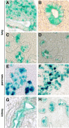
Injection of tamoxifen post-E7.5 results in epithelial-specific expression of β-galactosidase. In all images, epithelia is outlined in black dashed lines. A–D: In the E15.5 lung, injection at E5.5 (A) or E6.5 (B) labels mesenchymal cells, yet injections at E7.5 and later, shown here at E11.5 (C) or E12.5 (D), only label epithelia. Stage of harvesting: (A,B), E12.5; (C,D), E15.5. E,F: In the E12.5 pancreas, injection at E6.5 (E) leads to strong staining in the epithelia as well as in some scattered mesenchyme. Injection at E7.5 (F) is restricted to the epithelia. No mesenchymal staining was seen in any section examined. G,H: In the E15.5 kidney, injection at E11.5 (G) and E12.5 (H) are shown. All magnification at 10× except F at 20×. inj., injection.

References

    1. Ang SL, Rossant J. HNF-3β is essential for node and notochord formation in mouse development. Cell. 1994;78:561–574. - PubMed
    1. Apelqvist A, Ahlgren U, Edlund H. Sonic hedgehog directs specialised mesoderm differentiation in the intestine and pancreas. Curr Biol. 1997;7:801–804. - PubMed
    1. Arabzadeh A, Troy TC, Turksen K. Role of the Cldn6 cytoplasmic tail domain in membrane targeting and epidermal differentiation in vivo. Mol Cell Biol. 2006;26:5876–5887. - PMC - PubMed
    1. Batlle E, Sancho E, Francí C, Domínguez D, Monfar M, Baulida J, García de Herreros A. The transcription factor snail is a repressor of E-cadherin gene expression in epithelial tumour cells. Nat Cell Biol. 2000;2:84–89. - PubMed
    1. Belo JA, Bachiller D, Agius E, Kemp C, Borges AC, Marques S, Piccolo S, De Robertis EM. Cerberus-like is a secreted BMP and nodal antagonist not essential for mouse development. Genesis. 2000;26:265–270. - PubMed

Publication types

LinkOut - more resources