Local and remote lesions in horses subjected to small colon distension and decompression
- PMID: 18214165
- PMCID: PMC2117370
Local and remote lesions in horses subjected to small colon distension and decompression
Abstract
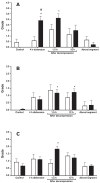
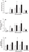
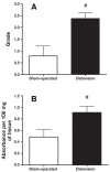
The purpose of this study was to observe and characterize colonic and lung lesions in horses subjected to experimental distension and decompression of the small colon. Sixteen healthy adult horses were divided into 2 groups: 9 horses that were subjected to distension of the small colon by means of a latex balloon surgically implanted in the lumen and inflated to a pressure of 40 mm Hg for 4 h, and 7 horses in which the balloon was implanted but not inflated. Colonic biopsy specimens were collected before balloon implantation, at the end of the period of obstruction, and 1.5 and 12 h after decompression and were examined for hemorrhage, edema, and neutrophil infiltration; myeloperoxidase (MPO) activity and hemoglobin concentration were measured as well. At the end of the experiment, lung samples were also collected and examined for neutrophil accumulation and MPO activity. The mucosa was not affected by luminal distension; lesions were restricted to the seromuscular layer. Neutrophil accumulation and edema were observed in the samples from both groups of horses but were greater in those from the distension group, in which there was also hemorrhage, fibrin deposition, and increased MPO activity in the seromuscular layer. Similarly, there was greater accumulation of neutrophils in the lung samples from the distension group than in those from the sham-operated group, as determined by histologic evaluation and MPO assay. These findings provide new evidence of reperfusion injury and a systemic inflammatory response, followed by remote lesions, in horses with intestinal obstruction.
Le but de cette étude était d’observer et de caractériser les lésions au côlon et au poumon chez des chevaux soumis à une distension et décompression expérimentale du petit côlon. Seize chevaux adultes ont été divisés en 2 groupes : 9 chevaux qui ont été soumis à une distension du petit côlon au moyen d’un ballon de latex implanté chirurgicalement dans la lumière et gonflé à une pression de 40 mm Hg pour 4 h, et 7 chevaux chez qui le ballon a été implanté mais non gonflé. Des spécimens de biopsie du côlon ont été prélevés avant l’implantation du ballon, à la fin de la période d’obstruction et 1,5 et 12 h après décompression et ont été observés pour la présence d’hémorragie, œdème, et infiltration de neutrophiles; l’activité de la myéloperoxidase (MPO) et la concentration d’hémoglobine ont également été mesurées. À la fin de l’expérience, des échantillons de poumon ont été prélevés et examinés pour l’accumulation de neutrophiles et l’activité de la MPO. La muqueuse n’était pas affectée par la distension de la lumière intestinale; les lésions étaient limitées à la couche séromusculaire. L’accumulation de neutrophiles et l’œdème étaient observés dans les échantillons provenant des deux groupes de chevaux mais étaient plus marqués chez les chevaux soumis à une distension, chez qui on observa également des hémorragies, la déposition de fibrine et une activité de la MPO augmentée dans la couche séromusculaire. De manière similaire, il y avait une plus grande accumulation de neutrophiles dans les échantillons de poumon provenant des chevaux du groupe ayant subit une distension que chez les chevaux du groupe témoin, tel que déterminé par l’évaluation histologique et les résultats des épreuves de la MPO. Ces résultats fournissent de nouvelles évidences des dommages de reperfusion et de réponse inflammatoire systémique, suivi de lésions à distance, chez des chevaux avec obstruction intestinale.
(Traduit par Docteur Serge Messier)
Figures


References
-
- Horne MM, Pascoe PJ, Ducharme NG, Barker IK, Grovum WL. Attempts to modify reperfusion injury of equine jejunal mucosa using dimethylsulfoxide, allopurinol, and intraluminal oxygen. Vet Surg. 1994;4:241–249. - PubMed
-
- Moore RM, Bertone AL, Muir WW, Stromberg PC, Beard WL. Histologic evidence of reperfusion injury in the large colon of horses after low-flow ischemia. Am J Vet Res. 1994;55:1434–1443. - PubMed
-
- Moore RM, Muir WW, Granger DN. Mechanisms of gastrointestinal ischemia-reperfusion injury and therapeutic interventions: a review and its implications in the horse. J Vet Intern Med. 1995;9:115–132. - PubMed
-
- Wilson DV, Patterson JS, Stick JA, Provost PJ. Histologic and ultrastructural changes after large-colon torsion, with and without use of a specific platelet-activating factor antagonist (WEB 2086) in ponies. Am J Vet Res. 1994;55:681–688. - PubMed
-
- Moore RM, Muir WW, Bertone AL, Beard WL, Stromberg PC. Effects of dimethyl sulfoxide, allopurinol, 21-aminosteroid U-74389G and manganese chloride on low-flow ischemia and reperfusion of the large colon in horses. Am J Vet Res. 1995;56:671–687. - PubMed
Publication types
MeSH terms
Substances
LinkOut - more resources
Full Text Sources
Medical
Research Materials
Miscellaneous
