. 2008 Apr;17(4):600-9.
doi: 10.1007/s00586-008-0599-8. Epub 2008 Jan 24.

En bloc spondylectomy in malignant tumors of the spine

Affiliations

En bloc spondylectomy in malignant tumors of the spine

Ulf Liljenqvist et al. Eur Spine J. 2008 Apr.

Abstract

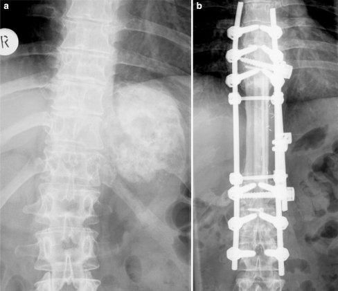
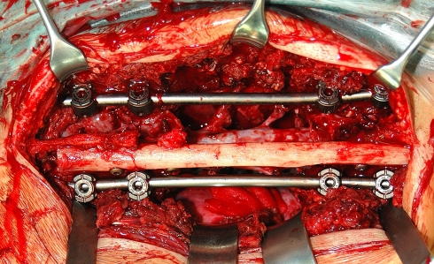
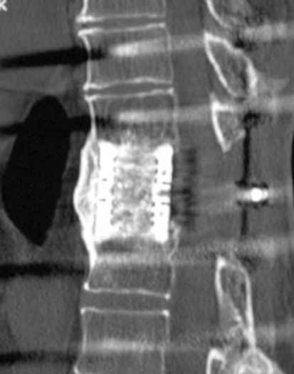
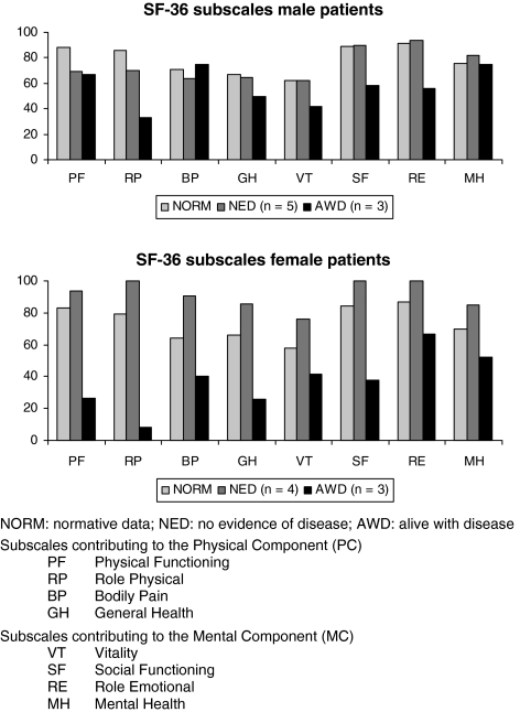
En bloc spondylectomy is a technique that enables wide or marginal resection of malignant lesions of the spine. Both all posterior techniques as well as combined approaches are reported. Aim of the present study was to analyse the results of 21 patients with malignant lesions of the spine, all treated with en bloc excision in a combined posteroanterior (n = 19) or all posterior approach (n = 2). Twenty-one consecutive patients, operated between 1997 and 2005, were included into this retrospective study. Thirteen patients had primary malignant lesions, eight patients had solitary metastases, all located in the thoracolumbar spine. There were 16 single level, three two-level, one three-level and one four-level spondylectomy. The patients were followed clinically and radiographically (including CT studies) with an average follow-up of 4 years. Out of 11 patients with primary Ewing or osteosarcoma seven patients are alive without any evidence of disease. One patient died after 5 years from other causes and three are alive with evidence of disease. Latter had either a poor histologic response to the preoperative chemotherapy (n = 2) or an intralesional resection (n = 1). All three patients with solitary spinal metastases of Ewing or osteosarcoma died of the disease. Five patients with solitary metastases of mainly hypernephroma are alive. In total, six resections were intralesional, mainly due to large intraspinal tumor masses, with two patients having had previous surgery. In the remaining cases, wide (n = 10) or marginal (n = 5) resection was accomplished. There were one pseudarthrosis requiring extension of the fusion and two cases with local recurrences and repeated excisional surgery. At follow-up CT studies, all cages were fused. Health related quality of life analysis (SF-36) revealed only slightly decreased physical component and normal mental component scores compared to normals in those patients with no evidence of disease. En bloc spondylectomy enables wide or marginal resection of malignant lesions of the spine in most cases with acceptable morbidity. Intralesional resection, poor histologic response, and solitary spinal metastases of Ewing and osteosarcoma are associated with a poor prognosis.


Figures

Fig. 1
Fig. 1
a Illustration of tumor involvement of one pedicle. b The non affected, healthy posterior elements are resected including the pedicle in order to create a corridor to release the dural tube
Fig. 2
Fig. 2
a–c 16-year-old male with Ewing’s sarcoma of L2 (patient number 6) and involvement of the left pedicle and transverse process. d Intraoperative view after resection of the healthy posterior structures to the base of the right pedicle with release of the right L2 nerve root. e and f Posterior and anterior view after en bloc spondylectomy. g Anterior column reconstruction with expandable titanium cage. h and i The cortical graft is trimmed and placed pressfit inbetween the spinous processes and covered with bone chips. j and k Macrospecimen and macrosection of L2 demonstrating clear margins. l and m Postoperative X-rays. n and o CT scans 3 years postop. showing bony fusion of both the cage and the posterior cortical graft
Fig. 2
Fig. 2
a–c 16-year-old male with Ewing’s sarcoma of L2 (patient number 6) and involvement of the left pedicle and transverse process. d Intraoperative view after resection of the healthy posterior structures to the base of the right pedicle with release of the right L2 nerve root. e and f Posterior and anterior view after en bloc spondylectomy. g Anterior column reconstruction with expandable titanium cage. h and i The cortical graft is trimmed and placed pressfit inbetween the spinous processes and covered with bone chips. j and k Macrospecimen and macrosection of L2 demonstrating clear margins. l and m Postoperative X-rays. n and o CT scans 3 years postop. showing bony fusion of both the cage and the posterior cortical graft
Fig. 3
Fig. 3
Preoperative and 6 years follow-up X-rays of patient number 8 with a large osteosarcoma involving T9–12. En bloc spondylectomy of four vertebrae with anterior fibula reconstruction and single rod instrumentation and posterior fibula fusion and instrumentation T6–L3
Fig. 4
Fig. 4
Intraoperative view of a two-level en bloc spondylectomy with posterior fixation of an allogenic fibula strut graft secured with titanium screws to the spinous processes
Fig. 5
Fig. 5
CT scan 42 months post en bloc spondylectomy T11 for solitary metastasis of a hypernephroma (patient number 19) demonstrating solid bony growth through the cage and continuous bony bridging anterior to the cage
Fig. 6
Fig. 6
Health related quality of life data of our male and female patients

References

    1. Bielack SS, Kempf-Bielack B, Delling G, Exner G, Flege S, et al. Prognostic factors in high-grade osteosarcoma of the extremities or trunk: an analysis of 1702 patients treated on neoadjuvant cooperative osteosarcoma study group protocols. J Clin Oncol. 2002;1:776–790. doi: 10.1200/JCO.20.3.776. - DOI - PubMed
    1. Boriani S, Biagini R, DeLure F. En bloc resections of bone tumors of the thoracolumbar spine. Spine. 1996;21:1927–1931. doi: 10.1097/00007632-199608150-00020. - DOI - PubMed
    1. Boriani S, Chevally F, Weinstein J. Chordoma of the spine above the sacrum. Spine. 1996;21:1569–1577. doi: 10.1097/00007632-199607010-00017. - DOI - PubMed
    1. Boriani S, Weinstein J, Biagini R. Primary bone tumors of the spine. Spine. 1997;22:1036–1044. doi: 10.1097/00007632-199705010-00020. - DOI - PubMed
    1. Ellert U, Bellach B. German national health survey 1998—description of an update random sampling test. Gesundheitswesen. 1999;61:S184–S190. - PubMed

MeSH terms