. 2008 Jan;6(1):e7.
doi: 10.1371/journal.pbio.0060007.

Specific targeting of a plasmodesmal protein affecting cell-to-cell communication

Affiliations

Specific targeting of a plasmodesmal protein affecting cell-to-cell communication

Carole L Thomas et al. PLoS Biol. 2008 Jan.

Abstract

Plasmodesmata provide the cytoplasmic conduits for cell-to-cell communication throughout plant tissues and participate in a diverse set of non-cell-autonomous functions. Despite their central role in growth and development and defence, resolving their modus operandi remains a major challenge in plant biology. Features of protein sequences and/or structure that determine protein targeting to plasmodesmata were previously unknown. We identify here a novel family of plasmodesmata-located proteins (called PDLP1) whose members have the features of type I membrane receptor-like proteins. We focus our studies on the first identified type member (namely At5g43980, or PDLP1a) and show that, following its altered expression, it is effective in modulating cell-to-cell trafficking. PDLP1a is targeted to plasmodesmata via the secretory pathway in a Brefeldin A-sensitive and COPII-dependent manner, and resides at plasmodesmata with its C-terminus in the cytoplasmic domain and its N-terminus in the apoplast. Using a deletion analysis, we show that the single transmembrane domain (TMD) of PDLP1a contains all the information necessary for intracellular targeting of this type I membrane protein to plasmodesmata, such that the TMD can be used to target heterologous proteins to this location. These studies identify a new family of plasmodesmal proteins that affect cell-to-cell communication. They exhibit a mode of intracellular trafficking and targeting novel for plant biology and provide technological opportunities for targeting different proteins to plasmodesmata to aid in plasmodesmal characterisation.


Conflict of interest statement

Competing interests. The authors have declared that no competing interests exist.

Figures

Figure 1
Figure 1. PDLP1a Targeting to Plasmodesmata
(A) shows a cartoon of a plasmodesma. The plasmodesma is seen as a PM-lined channel through the cell wall containing a central ER-derived rod-shaped desmotubule (DT). The location of possible proteinaceous components (arrows), as visualised by electron microscopy [6], in the symplastic channel is illustrated. (B–D) show subcellular location of PDLP1a:GFP following transgenic expression in Arabidopsis. Following expression of PDLP1a:GFP under the control of the CaMV 35S promoter (B) or the PDLP1a native promoter (C), optical sections through the lower epidermis show the targeting of PDLP1a to plasmodesmata. The upper images show the typical punctate distribution of plasmodesmata along the cell wall of adjoining cells. The lower images show an overlay of the fluorescence on the bright field image of the same area to identify the location of the cell walls. (D and E) PDLP1a:GFP remained at punctate structures within the cell wall after plasmolysis, as shown for epidermal cells (D), and on the adjoining wall between adjacent spongy mesophyll cells (E). An overlay of the fluorescence on the bright field image of the same area is shown; asterisks (*) demarcate the retracted protoplast, and arrows identify PDLP1a:GFP retained on the cell wall. Bar indicates 10 μm.
Figure 2
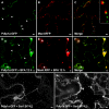
Figure 2. PDLP1a Resides in Plasmodesmata
Following coexpression of PDLP1a:GFP (A and D) and TMV MP:RFP (E), or chemical staining of callose using aniline blue (B), colocation of PDLP1a with either marker (C and F) confirmed its localisation to plasmodesmata. Bar indicates 5 μm.
Figure 3
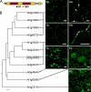
Figure 3. Organisation, Phylogenetic Analysis, and Subcellular Localisation of PDLP1a and Its Homologues
The domain structure of PDLP1a is shown in (A). The signal peptide (SP; black box) and the TMD (grey box) were predicted using SignalP 3.0 and TMHMM 2.0, respectively. The TMD is followed by a short cytoplasmic tail. The DUF26 domain (Pfam PF01657) is shown in red. Protein homology searches using the PDLP1a amino acid sequence, followed by phylogenetic analysis (B), revealed the presence of two clades within the closely related PDLP1 family; the gene for PDLP1a is boxed. All of these genes, in addition to representatives of more distantly related DUF26 proteins, were cloned and expressed trangenically in Arabidopsis from the CaMV 35S promoter. Optical sections through epidermal (Ep) and spongy mesophyll (Me) tissues of transgenic Arabidopsis expressing representatives of the two families of PDLP1a homologs (At2g33330/clade 1 and At2g01660/clade 2) show punctate labelling at all areas of cell–cell contact (some are marked with arrows). Optical sections through the epidermis of transgenic Arabidopsis expressing a 2xDUF26 protein lacking a TMD (encoded by At5g48540) shows fluorescent apoplastic bodies, whereas expression of a GPI-anchored 2xDUF26 relative (encoded by At1g63580) shows PM labelling; bright background fluorescence from stomata is also evident. The least-related 2xDUF26 with a TMD and a C-terminal kinase domain (encoded by At4g23140) also shows PM labelling. Bars indicate 10 μm .
Figure 4
Figure 4. PDLP1a Membrane Topology
(A) shows a schematic presentation of the expression cassettes for PDLP1a C-terminal fusions with YN and YC of YFP. N. benthamiana were agro-infiltrated with agrobacteria containing 35S::PDLP1a:YN or 35S::PDLP1a:YC and the corresponding counterpart (pLH::YN, pLH::YC, or pLH::YN-ER), and 35S::HCPro. (B) Five days post-inoculation, fluorescence was analysed by confocal microscopy confirming YFP fluorescence reconstitution in cells coexpressing 35S::YN and 35S::YC, 35S::YN-ER and 35S::YC-ER, 35S::PDLP1a:YN and 35S::YC, and 35S:PDLP1a:YC and 35S::YN, but not in cells coexpressing 35S::YN-ER and 35S::YC or 35S::PDLP1a:YC and 35S::YN-ER. (C) Bimolecular fluorescence confirmed the predicted orientation of PDLP1a as a type I membrane protein. Bars = 20 μm
Figure 5
Figure 5. Altered Expression of PDLP1 Changes Plasmodesmal Trafficking
PDLP1 KO lines (A) or PDLP1a:HA overexpressing lines (B) were assessed for plasmodesmal trafficking potential by counting the number of GFP recipient cells surrounding primary bombardment sites. From three double KO lines, only At2g33330×At1g04520 and At5g43980×At2g33330 showed altered GFP diffusion. (Data for At2g33330×At1g04520 were obtained from two independent experiments and 96 bombardment sites each for wild-type (WT) and KO lines (p ≤ 0.05); data for At5g43980×At2g33330 were obtained from three independent experiments and 57 and 93 bombardment sites for WT and KO lines, respectively (p ≤ 0.01). (B) Transgenic Arabidopsis plants expressing 35S::PDLP1a:HA (or 35S::PDLP1a:GFP; unpublished data) exhibited a dwarf phenotype (right, upper picture) when compared with nontransformed (NT) plants. This dwarf phenotype showed a positive correlation with transgene copy number and protein accumulation, assessed by western analysis with anti-HA antibody (right, lower picture). Bombardment of the less-dwarfed hemizygous plants (Hemi; left) showed a dramatic reduction in GFP diffusion (data collected from two independent experiments and 70 and 114 bombardment sites for the NT and hemizygous plants, respectively; p ≤ 0.0001). Homo, homozygous. Bars indicate the standard error of the mean.
Figure 6
Figure 6. PDLP1a Utilises the Secretory Pathway for Delivery to Plasmodesmata
N. benthamiana leaves were agro-infiltrated with a construct expressing 35S::PDLP1a:GFP (A to H) and either a construct expressing a Golgi marker (35S::ManI:RFP; [A to F]) or a construct expressing the Sar1 inhibitory mutant variant Sar1[H74L]:RFP that blocks ER to Golgi transport (G and H). (A–C) Cells expressing 35S::PDLP1a:GFP and 35S::ManI:RFP showed discrete punctate structures characteristic of plasmodesmata (arrowheads), and Golgi stacks, respectively. Inset, high magnification of the boxed region in (C) confirms the discrete nature of the two subcellular locations. (D–F) Treatment with BFA led to a severe reduction in number of cell wall–associated PDLP1a:GFP-labeled punctate structures ([D], compare to [A], arrowheads), and the near complete loss of individual Golgi stacks ([B], compare to [E]), and concomitantly in the formation of typical BFA compartments (asterisks) in which both PDLP1a:GFP and ManI:RFP colocalized. (G and H) Similar phenomenon was observed upon coexpression of PDLP1a:GFP and the GTP-restricted mutant Sar1[H74L]. In median section (G), PDLP1a:GFP formed large aggregates in the nuclear vicinity that resembled the BFA compartment. Fluorescence extended also all around the cells and within cytoplasmic strands (G). In the cortex of the same cells (H), PDLP1a:GFP was found to be located in a typical ER polygonal network that was better seen upon higher magnification (inset, 3× magnification of the boxed region in [H]). Coexpression of the Sar1[H74L] and PDLP1a constructs in the same cell was confirmed from their respective fluorescent tags. Scale bars indicate 5 μm.
Figure 7
Figure 7. Mutation Analysis of the Transmembrane Domain and Cytoplasmic Tail of PDLP1a
(A and B) Multiple sequence alignment showing homology in the TMD within clade 1 of the PDLP1 family and greater variation in the cytoplasmic tail (CT) (A); the gene for PDLP1a is boxed. Deletions were made in the TMD and cytoplasmic tail (CT) of PDLP1a (B). (C) After transgenic expression in Arabidopsis, PDLP1aΔC:GFP deleted for the CT targeted to punctate structures in the cell wall. (D) In contrast, transgenic expression to produce PDLP1aΔC+:GFP deleted for the CT plus the three amino acids (LVL; underlined) at the end of the TMD, abolished punctate labelling in the cell wall and resulted in targeting to the ER. Bars indicate 10 μm.
Figure 8
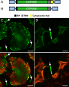
Figure 8. The TMD of PDLP1a Is Sufficient to Target Foreign Proteins to Plasmodesmata
(A and B) Chimeric constructs consisting of the signal peptide from PDLP1a, followed by the coding sequence for YFP (citrine) fused to the TMD and CT of PDLP1a (A), or the TMD alone (B), were constructed and expressed. (C and D) Upon plasmolysis of spongy mesophyll cells, the chimeric protein remained in the cell wall at the areas of cell–cell contact for both YFP:TMD+CT (C) and YFP:TMD (D) proteins. The former also showed accumulation of fluorescent bodies within the cytoplasm. Overlays of the YFP fluorescence and red chloroplast autofluorescence (bottom) images clearly show YFP labelling retained after plasmolysis at the points of cell-to-cell contact (arrows). Bars indicate 10 μm.

References

    1. Kurata T, Okada K, Wada T. Intercellular movement of transcription factors. Curr Opin Plant Biol. 2005;8:600–605. - PubMed
    1. Lee J-Y, Colinas J, Wang JY, Mace D, Ohler U, et al. Transcriptional and posttranscriptional regulation of transcription factor expression in Arabidopsis roots. Proc Natl Acad Sci U S A. 2006;103:6055–6060. - PMC - PubMed
    1. Dunoyer P, Himber C, Voinnet O. DICER-LIKE 4 is required for RNA interference and produces the 21-nucleotide small interfering RNA component of the plant cell-to-cell silencing signal. Nat Genet. 2005;37:1356–1360. - PubMed
    1. Lucas WJ. Plant viral movement proteins: agents for cell-to-cell trafficking of viral genomes. Virology. 2006;344:169–184. - PubMed
    1. Kankanala P, Czymmek K, Valent B. Roles for rice membrane dynamics and plasmodesmata during biotrophic invasion by the blast fungus. Plant Cell. 2007;19:706–724. - PMC - PubMed

Publication types

MeSH terms

Substances