. 2007 Dec;3(12):e199.
doi: 10.1371/journal.ppat.0030199.

A two-phase innate host response to alphavirus infection identified by mRNP-tagging in vivo

Affiliations

A two-phase innate host response to alphavirus infection identified by mRNP-tagging in vivo

Jennifer L Konopka et al. PLoS Pathog. 2007 Dec.

Abstract

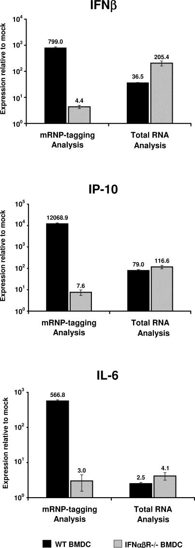
A concept fundamental to viral pathogenesis is that infection induces specific changes within the host cell, within specific tissues, or within the entire animal. These changes are reflected in a cascade of altered transcription patterns evident during infection. However, elucidation of this cascade in vivo has been limited by a general inability to distinguish changes occurring in the minority of infected cells from those in surrounding uninfected cells. To circumvent this inherent limitation of traditional gene expression profiling methods, an innovative mRNP-tagging technique was implemented to isolate host mRNA specifically from infected cells in vitro as well as in vivo following Venezuelan equine encephalitis virus (VEE) infection. This technique facilitated a direct characterization of the host defense response specifically within the first cells infected with VEE, while simultaneous total RNA analysis assessed the collective response of both the infected and uninfected cells. The result was a unique, multifaceted profile of the early response to VEE infection in primary dendritic cells, as well as in the draining lymph node, the initially targeted tissue in the mouse model. A dynamic environment of complex interactions was revealed, and suggested a two-step innate response in which activation of a subset of host genes in infected cells subsequently leads to activation of the surrounding uninfected cells. Our findings suggest that the application of viral mRNP-tagging systems, as introduced here, will facilitate a much more detailed understanding of the highly coordinated host response to infectious agents.


Conflict of interest statement

Competing interests. The authors have declared that no competing interests exist.

Figures

Figure 1
Figure 1. VEE Replicon Constructs
The VEE replicon genome encodes the viral nonstructural genes, the authentic 5′ and 3′ UTR, as well as a cloning cassette downstream of the 26S subgenomic promoter for transgene insertion. Schematic representation of the replicon constructs used in this study are shown. (A) FLAG-PABP VRP, expressing an N-terminally FLAG-tagged version of PABP (FLAG epitope denoted by checkered shading). Expression of FLAG-PABP expressed from the VRP, as well as endogenous PABP, is shown by western blotting against the FLAG epitope or against PABP itself, from L929 cell lysates harvested at 6 and 12 hpi. (B) Schematic of GFP-VRP, expressing green fluorescent protein. All replicon particles used in this study were packaged in the wildtype (V3000) envelope.
Figure 2
Figure 2. The VRP mRNP-Tagging System
Flowchart illustrating the VRP mRNA-tagging method of isolating mRNA specifically from infected cells. (A) Cells are infected with FLAG-PABP VRP, delivering the unique epitope-tagged version of PABP only to infected cells. (B) At various times post-infection, cytoplasmic lysates are prepared, containing the cellular mRNP complexes. (C) Anti-FLAG antibody–coated agarose beads are added in excess to the lysate, co-immunoprecipitating the mRNA bound by FLAG-PABP, and thus fractionating the mRNA in the infected cells from the mRNA in the surrounding uninfected cells. (D) The immunoprecipitated mRNA-PABP complex is dissociated using proteinase K digestion, and the infected cell mRNA is isolated by standard RNA extraction and precipitation.
Figure 3
Figure 3. RNA Profile Comparison following High MOI Infection
Changes in host gene expression were assessed following VRP infection, using RNA isolated by three distinct methods. L929 cells (106) were mock treated or infected with FLAG-PABP VRP (MOI=5). At 6, 12, or 24 h, cellular RNA was harvested by three separate methods: (i) Total RNA from mock and VRP-infected cells was isolated using UltraSpec reagent. (ii) All mRNA bound to PABP in lysates prepared from mock and VRP-infected cultures were isolated by anti-PABP immunoprecipitation. (iii) mRNA from infected cells was specifically isolated by anti-FLAG immunoprecipitation, recovering FLAG-PABP bound RNA in VRP-infected lysates. The separate RNA populations served as input RNA in an RPA to analyze expression profiles of several host genes. The fold induction of (A) IRF-1 and (B) IFNβ are shown above, comparing the profiles generated from each isolation technique. For the mock references, RNA was analyzed from mock treated cells using the corresponding isolation technique (e.g., signal from infected total RNA was compared to mock total RNA, and infected IP RNA was compared to mock IP RNA). The data are a representative of two separate experiments, with each sample internally normalized to GAPDH signal.
Figure 4
Figure 4. Sensitivity of the VRP mRNP-Tagging System
To assess the level of sensitivity in the mRNP-tagging system, an in vitro experiment was performed to model the in vivo–like condition of a diluted infected cell population. At 6 hpi, cell lysates (1 ml) were prepared from 106 L929 cells that had been infected with FLAG-PABP VRP (MOI=5). The resulting lysates were mixed in decreasing ratios of infected lysate to mock lysate, to a total volume of 200 μl. Anti-FLAG immunoprecipitation was performed to isolate RNA from the infected cell portion of this mixed lysate. To assess the mRNA signal recovered from the infected cells, an RPA was used to detect several host mRNAs, two of which are shown above (IRF-1, GAPDH). Signal from infected cell RNA within the mixed lysate was detected in samples comprised of as little as 1% infected cell lysate (highlighted by the small dots), a value approximately equal to 2 × 10³ infected cells. Quantitation of the IRF-1 signal can be found as supplementary data (Table S1, Figure S2).
Figure 5
Figure 5. Infected Cell Gene Expression Profiles Generated by mRNP-Tagging versus FACS-Based Assays
The VRP mRNP-tagging approach and a FACS-based method of sorting infected cells were compared. L929 cells (1.5 × 10⁶) were infected with either GFP-VRP or FLAG-PABP VRP (MOI=0.2). Twelve hours after GFP-VRP infection, infected cells were sorted and recovered based on GFP expression via FACS. The recovered GFP-positive (infected) cells were lysed, and all PABP-bound host messages were isolated by anti-PABP immunoprecipitation. In parallel, 12 h after FLAG-PABP VRP infection, the mRNP-tagging assay was used to directly sort the mRNA from infected cells by anti-FLAG immunoprecipitation. To evaluate and compare host gene expression in the infected cell populations, two independent samples were analyzed for IFNβ, IP-10, and IRF-1 expression by Taqman real-time PCR. The results were normalized to GAPDH signal, compared to PABP-bound mRNA from mock infected cells, and averaged.
Figure 6
Figure 6. Role of Interferon Signaling in VRP-Induced Host Gene Expression in Infected versus Bystander BMDC
BMDC (106) generated from wildtype 129sv/ev mice (black bars) were infected with FLAG-PABP VRP at a low MOI (0.5). At 6 h, RNA was isolated from mock and VRP-infected BMDC by either 1) preparing cell lysates for isolation of FLAG-PABP-bound mRNA via anti-FLAG immunoprecipitation, or 2) using UltraSpec reagent to isolate total cellular RNA. The mRNP-tagging method specifically isolated mRNA from the minority of infected BMDC via the bound FLAG-PABP. Conversely, total RNA extraction was used to isolate cellular RNA from the entire VRP-infected BMDC culture, with the majority of the population being DCs that had not been infected. To examine the contribution of signaling through the IFNαβ receptor, the same analysis was carried out in BMDC derived from IFNαβR−/− mice (hatched bars). cDNA was generated from each RNA isolation, and assessed for changes in host gene expression by Taqman real-time PCR. Three independent samples were normalized to GAPDH signal and analyzed in comparison to mock infected BMDC: Infected cell anti-FLAG(PABP) signal was compared to mock PABP signal, and infected total RNA signal was compared to mock total RNA signal. The data are shown as the geometric mean, ± the standard error of the mean.
Figure 7
Figure 7. In Vivo, the Combination of Traditional Profiling and mRNP-Tagging Uncovers Dynamic Gene Expression Changes within the DLN
Adult female Balb/c mice were inoculated in both rear footpads with 106 IU of FLAG-PABP VRP. Mock treated animals were inoculated with diluent alone. At 6 and 9 h, the popliteal DLNs were removed and washed with PBS. To isolate RNA from the entire cellular population of the DLN, both DLNs were pooled, homogenized, and total RNA extracted. For isolation of mRNA specifically from the infected cells of the DLN, the mRNA tagging technique was used: Five DLNs were pooled per sample and homogenized in lysis buffer, followed by anti-FLAG immunoprecipitation. cDNA was synthesized from each RNA sample, and Taqman real-time PCR was performed against several target host genes. Two independent pools were analyzed from each group, with GAPDH serving as the internal housekeeping control gene. Infected cell anti-FLAG (PABP) signal was compared to mock PABP signal, and infected total RNA signal was compared to mock total RNA signal. The results from each independent pool are graphed side-by-side. Asterisks indicate samples from infected DLN that had no detectable signal following anti-FLAG immunoprecipitation. mRNP-tagging analysis in conjunction with traditional profiling reveals two distinct, comprehensive views of the response to infection within this target tissue in vivo.

Similar articles

Cited by

References

    1. Tenenbaum SA, Carson CC, Lager PJ, Keene JD. Identifying mRNA subsets in messenger ribonucleoprotein complexes by using cDNA arrays. Proc Natl Acad Sci U S A. 2000;97:14085–14090. - PMC - PubMed
    1. Tenenbaum SA, Lager PJ, Carson CC, Keene JD. Ribonomics: identifying mRNA subsets in mRNP complexes using antibodies to RNA-binding proteins and genomic arrays. Methods. 2002;26:191–198. - PubMed
    1. Deo RC, Bonanno JB, Sonenberg N, Burley SK. Recognition of polyadenylate RNA by the poly(A)-binding protein. Cell. 1999;98:835–845. - PubMed
    1. Bernstein P, Ross J. Poly(A), poly(A) binding protein and the regulation of mRNA stability. Trends Biochem Sci. 1989;14:373–377. - PubMed
    1. Burd CG, Dreyfuss G. Conserved structures and diversity of functions of RNA-binding proteins. Science. 1994;265:615–621. - PubMed

Publication types

MeSH terms