Review
. 2008:3:249-77.
doi: 10.1146/annurev.pathmechdis.3.121806.154311.

Pathogenesis of thrombotic microangiopathies

Affiliations
Review

Pathogenesis of thrombotic microangiopathies

X Long Zheng et al. Annu Rev Pathol. 2008.

Abstract

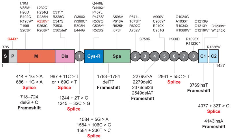
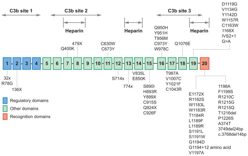
Profound thrombocytopenia and microangiopathic hemolytic anemia characterize thrombotic microangiopathy, which includes two major disorders: thrombotic thrombocytopenic purpura (TTP) and hemolytic uremic syndrome (HUS). TTP has at least three types: congenital or familial, idiopathic, and nonidiopathic. The congenital and idiopathic TTP syndromes are caused primarily by deficiency of ADAMTS13, owing to mutations in the ADAMTS13 gene or autoantibodies that inhibit ADAMTS13 activity. HUS is similar to TTP, but is associated with acute renal failure. Diarrhea-associated HUS accounts for more than 90% of cases and is usually caused by infection with Shiga-toxin-producing Escherichia coli (O157:H7). Diarrhea-negative HUS is associated with complement dysregulation in up to 50% of cases, caused by mutations in complement factor H, membrane cofactor protein, factor I or factor B, or by autoantibodies against factor H. The incomplete penetrance of mutations in either ADAMTS13 or complement regulatory genes suggests that precipitating events or triggers may be required to cause thrombotic microangiopathy in many patients.


Figures

Figure 1
Figure 1
Structure-function relationships of von Willebrand factor. Binding sites are indicated for collagen, factor VIII, platelet glycoprotein Ibα (GPIbα), and integrin αIIbβ III. The cleavage site (Tyr1605-Met1606) for ADAMTS13 is located at the central A2 domain of von Willebrand factor. The locations of intersubunit disulfide bonds (S-S) are shown.
Figure 2
Figure 2
The structure of ADAMTS13. The structural domains are indicated: signal peptide (S), propeptide (P), metalloprotease (M), disintegrin domain (Dis), first thrombospondin type 1 (TSP1) repeat, cysteine-rich domain (Cys-R), spacer domain (Spa), and two CUB domains (C1 and C2). The metalloprotease domain is the catalytic center that cleaves von Willebrand factor (VWF). The proximal carboxyl-terminal domains from Dis to Spa interact with the A2 domain of VWF. More distal carboxyl-terminal domains (TSP1 2–8) interact with VWF under fluid shear stress.
Figure 3
Figure 3
Location of the mutations found in patients with congenital thrombotic thrombocytopenic purpura. The point mutations that cause single amino acid substitutions and premature stop codons (X) are shown above the domain structure of ADAMTS13. The mutations that result in alternative splicing of ADAMTS13 mRNA or frameshifts are listed under the domain structure of ADAMTS13. Those mutants expressed as recombinant proteins in cell culture (*) cause defects in ADAMTS13 secretion (black) or catalytic activity (red ). S, signal peptide; P, propeptide; M, metalloprotease; Dis, disintegrin domain; 1, the first thrombospondin type 1 (TSP1) repeat; Cys-R, cysteine-rich domain; Spa, spacer domain; 2 through 8, the second to eighth TSP1 repeats; C1 and C2, the CUB domains 1 and 2.
Figure 4
Figure 4
Structure-function relationship of factor H and mutations found in patients with diarrhea-negative hemolytic uremic syndrome (D-HUS). The plasma protein factor H is composed of 20 consecutive short consensus repeat domains. The complement C3b and the heparin binding domains are indicated. Most of the mutations associated with D-HUS or familial HUS are clustered in the twentieth short consensus repeat domain.
Figure 5
Figure 5
The domain structure and mutations in membrane cofactor protein associated with diarrhea-negative hemolytic uremic syndrome (D-HUS). Membrane cofactor protein is an integral membrane protein composed of four short consensus repeat domains numbered 1 through 4, and O-glycosylated segments B and C rich in serine, threonine, and proline. Mutations associated with D-HUS or with familial HUS are indicated. An asterisk (*) indicates mutations that cause reduced levels of secretion.
Figure 6
Figure 6
Domain structure and mutations of factor I associated with diarrhea-negative hemolytic uremic syndrome. The heavy and light chains of factor I are linked by a disulfide bond (S-S). Most of the mutations in factor I are clustered in the serine protease domain. The mutations associated with reduced serum levels of factor I are marked with an asterisk (*).
Figure 7
Figure 7
The domain structure of complement factor B and mutations associated with diarrhea-negative hemolytic uremic syndrome. The schematic diagram of the factor B domain composition and the residues that participate in the Mg2+ binding are shown in panel a. The residues altered in patients with diarrhea-negative hemolytic uremic syndrome are shown in panel b.

References

    1. Moschcowitz E. Hyaline thrombosis of the terminal arterioles and capillaries: a hitherto undescribed disease. Proc NY Pathol Soc. 1924;24:21–24.
    1. Amorosi EL, Ultmann JE. Thrombocytopic purpura: report of 16 cases and review of the literature. Medicine. 1966;45:139–59.
    1. George JN, Gilcher RO, Smith JW, Chandler L, Duvall D, et al. Thrombotic thrombocytopenic purpura-hemolytic uremic syndrome: diagnosis and management. J Clin Apher. 1998;13:120–25. - PubMed
    1. George JN. How I treat patients with thrombotic thrombocytopenic purpura-hemolytic uremic syndrome. Blood. 2000;96:1223–29. - PubMed
    1. Bell WR, Braine HG, Ness PM, Kickler TS. Improved survival in thrombotic thrombocytopenic purpura-hemolytic uremic syndrome. Clinical experience in 108 patients. N Engl J Med. 1991;325:398–403. - PubMed

Publication types

MeSH terms

LinkOut - more resources