Comparative Study
. 2008 Jan;27(2):352-63.
doi: 10.1111/j.1460-9568.2008.06024.x.

Characterization of GABAergic neurons in rapid-eye-movement sleep controlling regions of the brainstem reticular formation in GAD67-green fluorescent protein knock-in mice

Affiliations
Comparative Study

Characterization of GABAergic neurons in rapid-eye-movement sleep controlling regions of the brainstem reticular formation in GAD67-green fluorescent protein knock-in mice

Ritchie E Brown et al. Eur J Neurosci. 2008 Jan.

Abstract

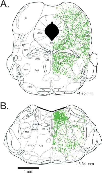
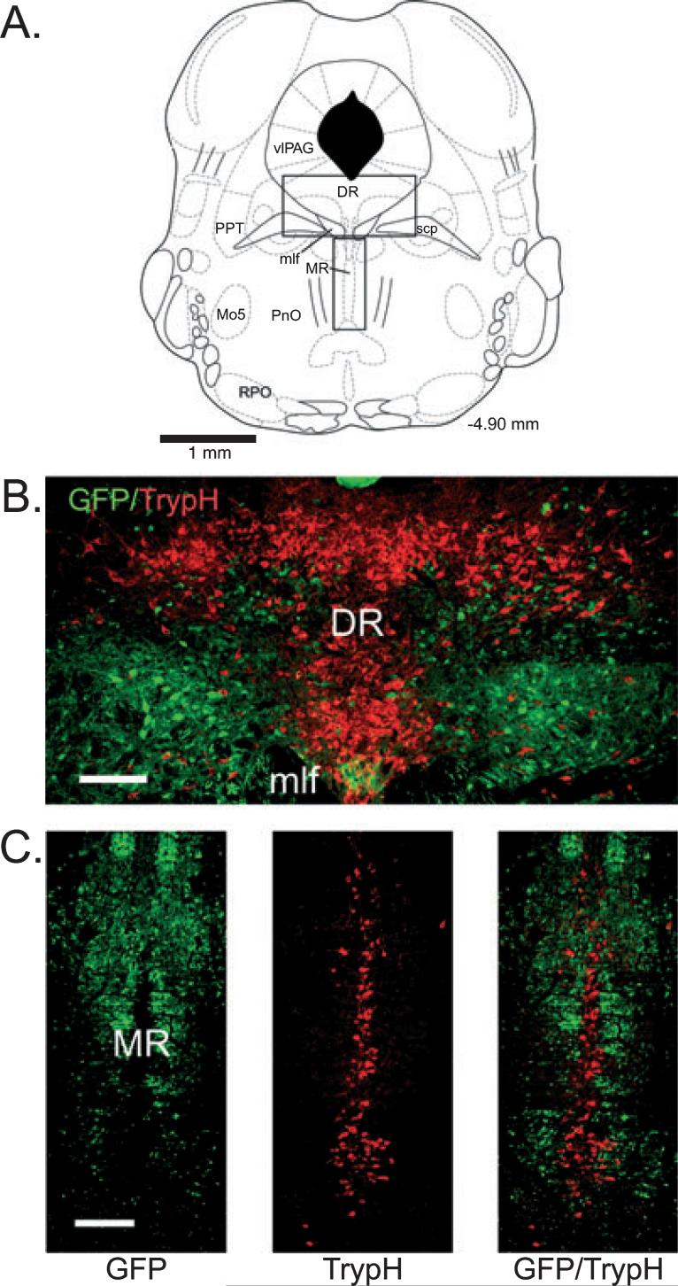
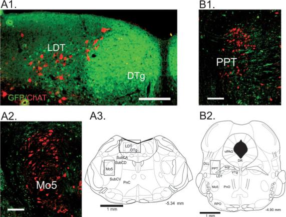
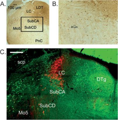
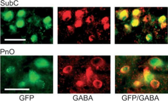
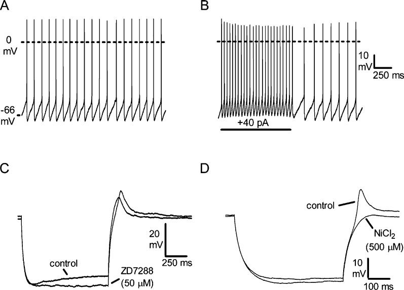
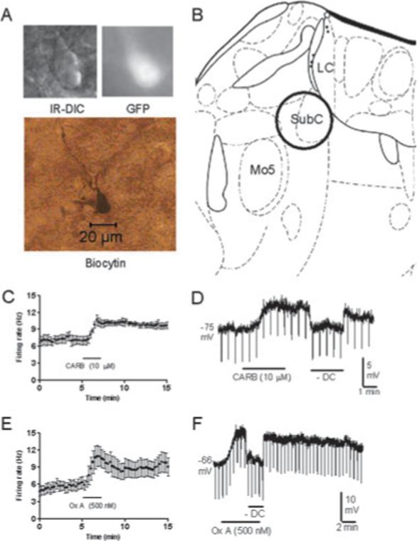
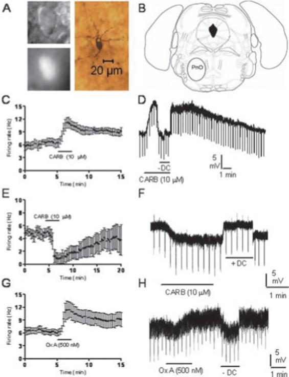
Recent experiments suggest that brainstem GABAergic neurons may control rapid-eye-movement (REM) sleep. However, understanding their pharmacology/physiology has been hindered by difficulty in identification. Here we report that mice expressing green fluorescent protein (GFP) under the control of the GAD67 promoter (GAD67-GFP knock-in mice) exhibit numerous GFP-positive neurons in the central gray and reticular formation, allowing on-line identification in vitro. Small (10-15 microm) or medium-sized (15-25 microm) GFP-positive perikarya surrounded larger serotonergic, noradrenergic, cholinergic and reticular neurons, and > 96% of neurons were double-labeled for GFP and GABA, confirming that GFP-positive neurons are GABAergic. Whole-cell recordings in brainstem regions important for promoting REM sleep [subcoeruleus (SubC) or pontine nucleus oralis (PnO) regions] revealed that GFP-positive neurons were spontaneously active at 3-12 Hz, fired tonically, and possessed a medium-sized depolarizing sag during hyperpolarizing steps. Many neurons also exhibited a small, low-threshold calcium spike. GFP-positive neurons were tested with pharmacological agents known to promote (carbachol) or inhibit (orexin A) REM sleep. SubC GFP-positive neurons were excited by the cholinergic agonist carbachol, whereas those in the PnO were either inhibited or excited. GFP-positive neurons in both areas were excited by orexins/hypocretins. These data are congruent with the hypothesis that carbachol-inhibited GABAergic PnO neurons project to, and inhibit, REM-on SubC reticular neurons during waking, whereas carbachol-excited SubC and PnO GABAergic neurons are involved in silencing locus coeruleus and dorsal raphe aminergic neurons during REM sleep. Orexinergic suppression of REM during waking is probably mediated in part via excitation of acetylcholine-inhibited GABAergic neurons.


Figures

Fig. 1
Fig. 1
Location of green fluorescent protein-positive neuronal perikarya in the brainstem of GAD67-GFP knock-in mice at the level of the dorsal raphe nucleus (A) and locus coeruleus (B). Numbers indicate the rostrocaudal location of the section with respect to Bregma. DLL, dorsolateral lemniscus; DMTg, dorsomedial tegmental area; DTg, dorsal tegmental nucleus of Gudden; DR, dorsal raphe nucleus; IC, inferior colliculus; LC, locus coeruelus; LDT, laterodorsal tegmentum; LPB, lateral parabrachial nucleus; LPT, lateral pontine tegmentum; mlf, medial longitudinal fasciculus; Mo5, motor trigeminal nucleus; MR, median raphe nucleus; PnC, pontine reticular nucleus, caudal part; PnO, pontine nucleus oralis; PPT, pedunculopontine tegmental area; RPO, rostral periolivary region; scp, superior cerebellar peduncle; SubCA, subcoeruleus nucleus, alpha part; SubCD, subcoeruleus nucleus, dorsal part; SubCV, subcoeruleus nucleus, ventral part; vlPAG, ventrolateral periaqueductal gray; VTg, ventral tegmental nucleus of Gudden.
Fig. 2
Fig. 2
Green fluorescent protein (GFP)-positive neurons have a complementary distribution to serotonergic neurons in the dorsal and median raphe. (A) Schematic indicating the locations (rectangles) of the dorsal raphe (DR) and median raphe (MR) regions depicted in B and C, respectively (adapted from Paxinos & Franklin, 2001). (B and C) Fluorescent images of GFP-positive neurons and tryptophan hydroxylase (TrypH)-positive (serotonergic) neurons labeled with streptavidin–Cy3 (red). Note that GFP-positive neurons are largely absent from midline areas where TrypH-positive neurons are most highly concentrated. For abbreviations, see legend for Fig. 1. Scale bars: 150 μm.
Fig. 3
Fig. 3
Relationship of green fluorescent protein (GFP)-positive neurons to cholinergic brainstem neurons. Fluorescent images of GFP-positive neurons and choline acetyltransferase (ChAT)-positive (cholinergic) neurons labeled with streptavidin–Cy3 (red). (A1) At the level of the caudal laterodorsal tegmental nucleus, numerous small GFP-positive neurons are densely packed in the dorsal tegmental nucleus of Gudden (DTg) and are intermingled with larger ChAT-positive neurons in the laterodorsal tegmentum (LDT). (A2) GFP-positive neurons form a ring around cholinergic motoneurons of the trigeminus (Mo5). (A3) Schematic indicating the locations (rectangles) of the regions depicted in A1 and A2 (adapted from Paxinos & Franklin, 2001). (B1) Small GFP-positive neurons are intermingled with larger ChAT-positive neurons in the pedunculopontine tegmental nucleus (PPT). GFP is also present within fibers in this area. (B2) Schematic indicating the location (rectangle) of the region depicted in B1 (adapted from Paxinos & Franklin, 2001). For abbreviations, see legend for Fig. 1. Scale bars: 150 μm.
Fig. 4
Fig. 4
Green fluorescent protein (GFP)-positive neurons are located within the subcoeruleus (SubC) area that is important for REM muscle atonia. (A) Location of the SubC area ventral to the locus coeruleus (LC). The slide was processed for anti-GFP staining revealed using diaminobenzidine (DAB). The area indicated by the rectangle is shown at higher power in B. A moderate number of round or triangular-shaped neurons (stained brown) can be seen in this area. (C) Fluorescent image of GFP-positive neurons and tyrosine hydroxylase (TyH)-positive (noradrenaline) neurons labeled with streptavidin–Cy3 (red) at the level of the main part of the LC. GFP-positive neurons are intermingled with scattered larger TyH-positive neurons ventral to the LC in the alpha (SubCA) and dorsal (SubCD) parts of the SubC. For abbreviations, see legend for Fig. 1. Scale bar: 200 μm.
Fig. 5
Fig. 5
Green fluorescent protein (GFP)-positive neurons contain GABA in the subcoeruleus (SubC) and pontine nucleus oralis (PnO) regions used for electrophysiological recordings. Left: Fluorescent images of GFP-positive neurons. Note that the fluorescence fills the entire cell, including the nucleus. Note also that punctate green fluorescence suggestive of axon terminals can be observed over the somata of several neurons (especially in the PnO). Middle: Fluorescent images of neurons stained for GABA/streptavidin–Cy3 (red). Note that the staining is cytoplasmic and avoids the nucleus. Right: Overlay of the green and red fluorescent images reveals that the GFP-positive neurons (green) in the SubC and PnO contain GABA (red), producing an orange color or orange with a green center (GFP in the nucleus). Scale bars: 25 μm.
Fig. 6
Fig. 6
Intrinsic membrane properties of subcoeruleus (SubC) and pontine nucleus oralis (PnO) green fluorescent protein (GFP)-positive neurons (n = 61). (A) Spontaneous action potential firing (scale same as in B). (B) Increased tonic firing in response to depolarizing current step. (C) Response to hyperpolarizing current injection (−130 pA) in the presence of tetrodotoxin (TTX, 0.5 μM), illustrating a medium-sized depolarizing sag during the step that is blocked by the H-current blocker ZD7288 (n = 7/7). Note that the rebound depolarization at the offset of the step is slightly increased in size by the block of Ih. (D) Response to a smaller hyperpolarizing current step (−100 pA) in a different neuron, illustrating a rebound depolarization that is blocked by the calcium channel antagonist NiCl2 (n = 9/9).
Fig. 7
Fig. 7
Pharmacological properties of green fluorescent protein (GFP)-positive neurons in the subcoeruleus (SubC). (A) Infrared differential interference contrast (IR-DIC) image, black-and-white image of GFP fluorescence, and post hoc biocytin stain (below) of the recorded neuron. (B) Location of the SubC in the mouse brainstem (adapted from Paxinos & Franklin, 2001). LC, locus coeruleus; Mo5, motor trigeminal nucleus. (C and D) Response of neurons to carbachol (CARB) in cell-attached mode (C, n = 5/5) and in whole-cell current-clamp mode in the presence of tetrodotoxin (D, n = 5/5). SubC GFP-positive neurons were excited by CARB. (E and F) Response of neurons to orexin A (Ox A) in cell-attached mode (E, n = 7/7) and in whole-cell current-clamp mode in the presence of tetrodotoxin (F, n = 5/5). SubC GFP-positive neurons were excited by Ox A. Downward deflections in D and F reflect hyperpolarizing current pulses used to monitor input resistance. Hyperpolarizing current injection (–DC) was used so as to compare input resistance at the baseline membrane potential.
Fig. 8
Fig. 8
Pharmacological properties of green fluorescent protein (GFP)-positive neurons in the pontine nucleus oralis (PnO). (A) Infrared differential interference contrast (IR-DIC) image, black-and-white image of GFP fluorescence, and post hoc biocytin stain (below) of the recorded neuron. (B) Location of the PnO in the mouse brainstem (adapted from Paxinos & Franklin, 2001). (C–F) Response of neurons to carbachol (CARB) in cell-attached mode (C and E) or in whole-cell current-clamp mode in the presence of tetrodotoxin (D and F). Different populations of PnO GFP-positive neurons were excited (C and D; n = 8/12, n = 7/11, respectively) or inhibited (E and F; n = 4/12, n = 4/11, respectively) by CARB. (E and F) Response of neurons to orexin A (Ox A) in cell-attached mode (G, n = 6/6) or in whole-cell current-clamp mode in the presence of tetrodotoxin (H, n = 9/9). PnO GFP-positive neurons were excited by Ox A. Downward deflections in D and F reflect hyperpolarizing current pulses used to monitor input resistance. Hyperpolarizing current injection (–DC) was used so as to compare input resistance at the baseline membrane potential. Note that the recording presented in H appears noisy due to a high frequency of spontaneous inhibitory synaptic potentials.

References

    1. Acuna-Goycolea C, Tamamaki N, Yanagawa Y, Obata K, van den Pol AN. Mechanisms of neuropeptide Y, peptide YY, and pancreatic polypeptide inhibition of identified green fluorescent protein-expressing GABA neurons in the hypothalamic neuroendocrine arcuate nucleus. J. Neurosci. 2005;25:7406–7419. - PMC - PubMed
    1. Asada H, Kawamura Y, Maruyama K, Kume H, Ding R, Ji FY, Kanbara N, Kuzume H, Sanbo M, Yagi T, Obata K. Mice lacking the 65 kDa isoform of glutamic acid decarboxylase (GAD65) maintain normal levels of GAD67 and GABA in their brains but are susceptible to seizures. Biochem. Biophys. Res. Commun. 1996;229:891–895. - PubMed
    1. Asada H, Kawamura Y, Maruyama K, Kume H, Ding RG, Kanbara N, Kuzume H, Sanbo M, Yagi T, Obata K. Cleft palate and decreased brain gamma-aminobutyric acid in mice lacking the 67-kDa isoform of glutamic acid decarboxylase. Proc. Natl Acad. Sci. USA. 1997;94:6496–6499. - PMC - PubMed
    1. Aston-Jones G, Zhu Y, Card JP. Numerous GABAergic afferents to locus ceruleus in the pericerulear dendritic zone: possible interneuronal pool. J. Neurosci. 2004;24:2313–2321. - PMC - PubMed
    1. Boissard R, Gervasoni D, Schmidt MH, Barbagli B, Fort P, Luppi PH. The rat ponto-medullary network responsible for paradoxical sleep onset and maintenance: a combined microinjection and functional neuroanatomical study. Eur. J. Neurosci. 2002;16:1959–1973. - PubMed

Publication types

Substances