. 2008 Jan 23:5:6.
doi: 10.1186/1742-4690-5-6.

Human endogenous retrovirus-FRD envelope protein (syncytin 2) expression in normal and trisomy 21-affected placenta

Affiliations

Human endogenous retrovirus-FRD envelope protein (syncytin 2) expression in normal and trisomy 21-affected placenta

André Malassiné et al. Retrovirology. .

Abstract

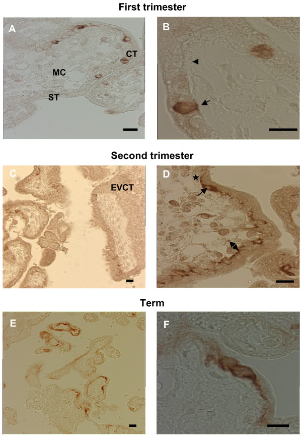
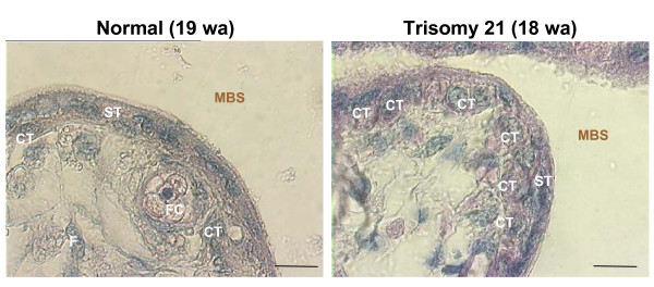
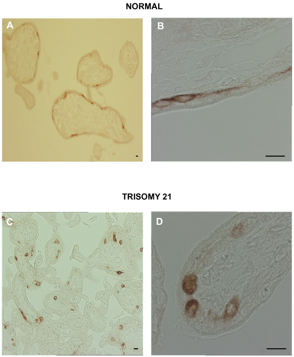
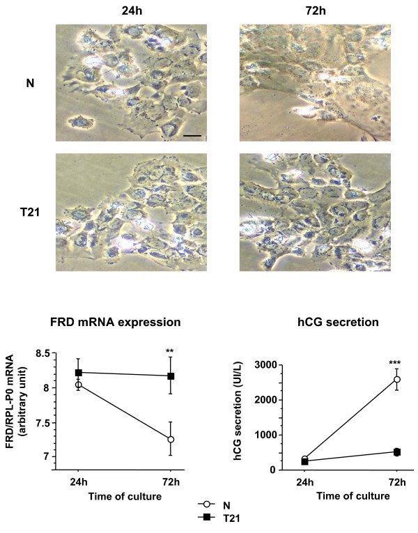
Human trophoblast expresses two fusogenic retroviral envelope proteins, the widely studied syncytin 1, encoded by HERV-W and the recently characterized syncytin 2 encoded by HERV-FRD. Here we studied syncytin 2 in normal and Trisomy 21-affected placenta associated with abnormal trophoblast differentiation. Syncytin 2 immunolocalization was restricted throughout normal pregnancy to some villous cytotrophoblastic cells (CT). During the second trimester of pregnancy, syncytin 2 was immunolocalized in some cuboidal CT in T21 placentas, whereas in normal placentas it was observed in flat CT, extending into their cytoplasmic processes. In vitro, CT isolated from normal placenta fuse and differentiate into syncytiotrophoblast. At the same time, syncytin 2 transcript levels decreased significantly with syncytiotrophoblast formation. In contrast, CT isolated from T21-affected placentas fused and differentiated poorly and no variation in syncytin 2 transcript levels was observed. Syncytin 2 expression illustrates the abnormal trophoblast differentiation observed in placenta of fetal T21-affected pregnancies.


Figures

Figure 1
Figure 1
A. In humans, at 10–12 weeks of pregnancy, the chorionic floating villi are in contact with the maternal blood in the maternal blood space (MBS). In these villi, cytotrophoblastic cells (CT) differentiate by fusion to generate the syncytiotrophoblast (ST). In the anchoring villi the cytotrophoblastic cells proliferate and invade the decidua (DC). The extravillous cytotrophoblastic cells (ECT) invade the lumen of uterine arteries (UA). FC: Fetal capillary; M: mesenchyme. B. Evolution of human floating chorionic villi. The chorionic villi, in direct contact with the maternal blood in the maternal blood space (MBS), consist of cytotrophoblastic cells (CT) and syncytiotrophoblast (ST) surrounding a core of mesenchymal cells including fetal capillaries (FC), fibroblasts (F) and Hofbauer cells (HC). BL: basal lamina.
Figure 2
Figure 2
Immunohistochemical analysis of syncytin 2 (HERV-FRD Env) in human placenta. Top panel. First trimester floating villi (8 weeks of pregnancy). A. Syncytin 2 was detected in some cytotrophoblastic cells (CT). No immunostaining was observed in the syncytiotrophoblast (ST) and in the mesenchymal core (MC). Scale bar = 10 μm. B. This large magnification allows to clearly establish the cytoplasmic localization of syncytin 2 immunostaining in a pair of cytotrophoblastic cells. At this gestational age the cytotrophoblast consists of a continuous single layer of cuboidal cells beneath the syncytiotrophoblast. Scale bar = 10 μm. Arrowhead: non labeled CT, arrow: positively stained CT. Middle panel. Second trimester placenta (16 weeks of pregnancy). C. Immunostaining with anti-syncytin2 antibody shows positive reactivity in some cytotrophoblastic cells. No syncytin 2 reactivity was detected in the extravillous trophoblast (ECT), in the syncytiotrophoblast and in the mesenchymal core. Scale bar = 10 μm. D. In this floating villi, syncytin 2 immunostaining was observed in the cytoplasm of some cytotrophoblastic cells (arrow), in their thin cytoplasmic processes (star) and at the level of the trophoblastic basal lamina (double head arrow). Scale bar = 10 μm. Bottom panel. Term placenta floating villi. E. Syncytin 2 was detected in the cytoplasm surrounding the nuclei of flat cytotrophoblastic cells and in their thin elongated cytoplasmic processes. Staining was absent from some villi. Scale bar = 10 μm. F. This large magnification allows to clearly establish the syncytin 2 immunostaining continuity within cytotrophoblasts between the cytoplasm surrounding the nuclei and that of the thin cytoplasmic processes. Scale bar = 10 μm.
Figure 3
Figure 3
Second trimester chorionic villi of normal (19 weeks of amenorrhea: wa) and trisomy 21 (18 wa) placentae. In normal placenta, a large amount of cytotrophoblastic cells (CT) have fused into a thin multinucleated syncytiotrophoblast (ST). In trisomy 21 placenta, many cuboidal cytotrophoblastic cells (CT) are still present beneath the syncytiotrophoblast (ST) increasing the thickness of the trophoblastic layer. Scale bar = 10 μm.
Figure 4
Figure 4
Immunohistochemical analysis of syncytin 2 (HERV-FRD Env) in age-matched second trimester (19 weeks) normal (upper panel) and T21-affected placentas (lower panel). Upper panel A. Immunostaining with anti-syncytin2 antibody showed positive reactivity in a fraction of elongated cytotrophoblastic cells. Scale bar = 10 μm. B. In this large magnification, syncytin 2 immunostaining was observed in the cytoplasm of cytotrophoblastic cells and in the thin cytoplasmic processes. Scale bar = 10 μm. Lower panel C. Syncytin 2 was detected in some cuboidal cytotrophoblastic cells. Scale bar = 10 μm. D. This high magnification shows the cytoplasmic localization of syncytin 2 immunostaining in several cytotrophoblastic cells. Scale bar = 10 μm.
Figure 5
Figure 5
Morphological differentiation (upper panel), real-time RT-PCR analysis of syncytin 2 (HERV-FRD env) transcripts (lower left panel) and hCG secretion (lower rigth panel) during in vitro culture of control and T21 trophoblastic cells. Cytotrophoblastic cells were purified from three distinct age matched (second trimester) control and T21-affected placentas and separately cultured. The cells were visualized under phase contrast light microscopy (Scale bar = 10 μm). At 72 h, normal cytotrophoblastic cells had fused resulting in the formation of a large syncytium containing numerous nuclei. In contrast, T21 cytotrophoblasts were still aggregated and had not fused. Total mRNA were extracted after 24 and 72 h of culture. Data are expressed as the level of syncytin 2 mRNA normalized to that of RPLP0 mRNA. HCG secretion into the culture medium was measured at the indicated times, in normal (N) and T21-affected cell cultures. Results are the means ± SEM of triplicate dishes from three different cultures.

References

    1. de Parseval N, Heidmann T. Human endogenous retroviruses: from infectious elements to human genes. Cytogenet Genome Res. 2005;110:318–332. doi: 10.1159/000084964. - DOI - PubMed
    1. Lander ES, Linton LM, Birren B, Nusbaum C, Zody MC, Baldwin J, Devon K, Dewar K, Doyle M, FitzHugh W, Funke R, Gage D, Harris K, Heaford A, Howland J, Kann L, Lehoczky J, LeVine R, McEwan P, McKernan K, Meldrim J, Mesirov JP, Miranda C, Morris W, Naylor J, Raymond C, Rosetti M, Santos R, Sheridan A, Sougnez C, Stange-Thomann N, Stojanovic N, Subramanian A, Wyman D, Rogers J, Sulston J, Ainscough R, Beck S, Bentley D, Burton J, Clee C, Carter N, Coulson A, Deadman R, Deloukas P, Dunham A, Dunham I, Durbin R, French L, Grafham D, Gregory S, Hubbard T, Humphray S, Hunt A, Jones M, Lloyd C, McMurray A, Matthews L, Mercer S, Milne S, Mullikin JC, Mungall A, Plumb R, Ross M, Shownkeen R, Sims S, Waterston RH, Wilson RK, Hillier LW, McPherson JD, Marra MA, Mardis ER, Fulton LA, Chinwalla AT, Pepin KH, Gish WR, Chissoe SL, Wendl MC, Delehaunty KD, Miner TL, Delehaunty A, Kramer JB, Cook LL, Fulton RS, Johnson DL, Minx PJ, Clifton SW, Hawkins T, Branscomb E, Predki P, Richardson P, Wenning S, Slezak T, Doggett N, Cheng JF, Olsen A, Lucas S, Elkin C, Uberbacher E, Frazier M, Gibbs RA, Muzny DM, Scherer SE, Bouck JB, Sodergren EJ, Worley KC, Rives CM, Gorrell JH, Metzker ML, Naylor SL, Kucherlapati RS, Nelson DL, Weinstock GM, Sakaki Y, Fujiyama A, Hattori M, Yada T, Toyoda A, Itoh T, Kawagoe C, Watanabe H, Totoki Y, Taylor T, Weissenbach J, Heilig R, Saurin W, Artiguenave F, Brottier P, Bruls T, Pelletier E, Robert C, Wincker P, Smith DR, Doucette-Stamm L, Rubenfield M, Weinstock K, Lee HM, Dubois J, Rosenthal A, Platzer M, Nyakatura G, Taudien S, Rump A, Yang H, Yu J, Wang J, Huang G, Gu J, Hood L, Rowen L, Madan A, Qin S, Davis RW, Federspiel NA, Abola AP, Proctor MJ, Myers RM, Schmutz J, Dickson M, Grimwood J, Cox DR, Olson MV, Kaul R, Raymond C, Shimizu N, Kawasaki K, Minoshima S, Evans GA, Athanasiou M, Schultz R, Roe BA, Chen F, Pan H, Ramser J, Lehrach H, Reinhardt R, McCombie WR, de la Bastide M, Dedhia N, Blocker H, Hornischer K, Nordsiek G, Agarwala R, Aravind L, Bailey JA, Bateman A, Batzoglou S, Birney E, Bork P, Brown DG, Burge CB, Cerutti L, Chen HC, Church D, Clamp M, Copley RR, Doerks T, Eddy SR, Eichler EE, Furey TS, Galagan J, Gilbert JG, Harmon C, Hayashizaki Y, Haussler D, Hermjakob H, Hokamp K, Jang W, Johnson LS, Jones TA, Kasif S, Kaspryzk A, Kennedy S, Kent WJ, Kitts P, Koonin EV, Korf I, Kulp D, Lancet D, Lowe TM, McLysaght A, Mikkelsen T, Moran JV, Mulder N, Pollara VJ, Ponting CP, Schuler G, Schultz J, Slater G, Smit AF, Stupka E, Szustakowski J, Thierry-Mieg D, Thierry-Mieg J, Wagner L, Wallis J, Wheeler R, Williams A, Wolf YI, Wolfe KH, Yang SP, Yeh RF, Collins F, Guyer MS, Peterson J, Felsenfeld A, Wetterstrand KA, Patrinos A, Morgan MJ, de Jong P, Catanese JJ, Osoegawa K, Shizuya H, Choi S, Chen YJ. Initial sequencing and analysis of the human genome. Nature. 2001;409:860–921. doi: 10.1038/35057062. - DOI - PubMed
    1. de Parseval N, Lazar V, Casella JF, Benit L, Heidmann T. Survey of human genes of retroviral origin: identification and transcriptome of the genes with coding capacity for complete envelope proteins. J Virol. 2003;77:10414–10422. doi: 10.1128/JVI.77.19.10414-10422.2003. - DOI - PMC - PubMed
    1. Blaise S, de Parseval N, Benit L, Heidmann T. Genomewide screening for fusogenic human endogenous retrovirus envelopes identifies syncytin 2, a gene conserved on primate evolution. Proc Natl Acad Sci U S A. 2003;100:13013–13018. doi: 10.1073/pnas.2132646100. - DOI - PMC - PubMed
    1. Blond JL, Lavillette D, Cheynet V, Bouton O, Oriol G, Chapel-Fernandes S, Mandrand B, Mallet F, Cosset FL. An envelope glycoprotein of the human endogenous retrovirus HERV-W is expressed in the human placenta and fuses cells expressing the type D mammalian retrovirus receptor. J Virol. 2000;74:3321–3329. doi: 10.1128/JVI.74.7.3321-3329.2000. - DOI - PMC - PubMed

Publication types