

Molecular basis of telaprevir resistance due to V36 and T54 mutations in the NS3-4A protease of the hepatitis C virus
- PMID: 18215275
- PMCID: PMC2395260
- DOI: 10.1186/gb-2008-9-1-r16
Molecular basis of telaprevir resistance due to V36 and T54 mutations in the NS3-4A protease of the hepatitis C virus
Abstract
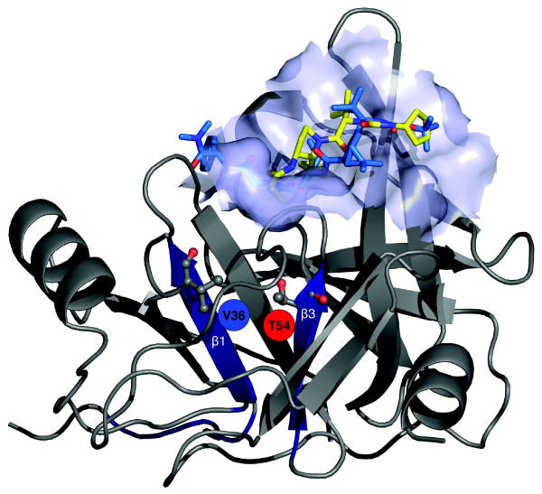
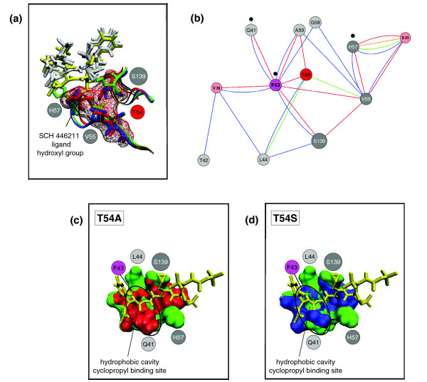
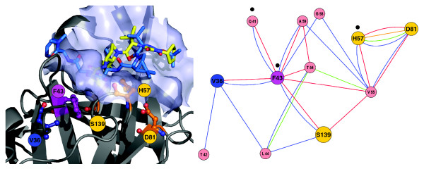
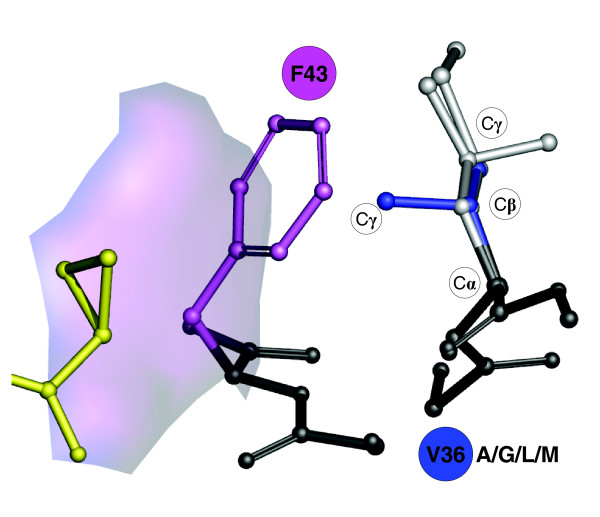
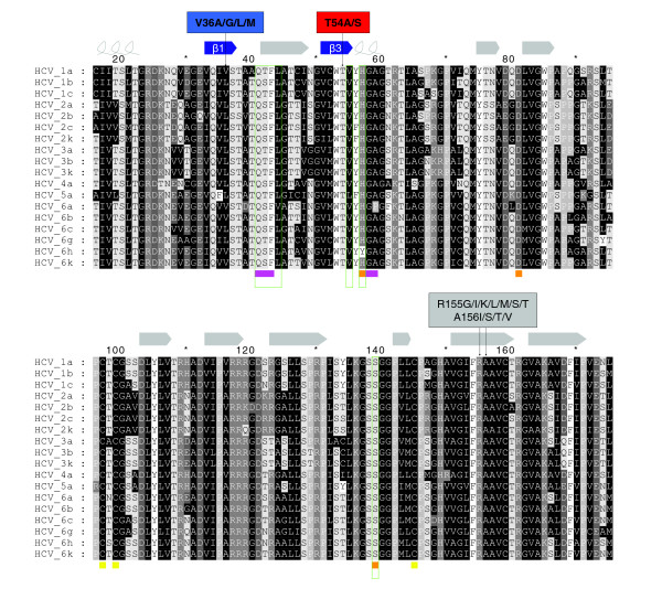
Background: The inhibitor telaprevir (VX-950) of the hepatitis C virus (HCV) protease NS3-4A has been tested in a recent phase 1b clinical trial in patients infected with HCV genotype 1. This trial revealed residue mutations that confer varying degrees of drug resistance. In particular, two protease positions with the mutations V36A/G/L/M and T54A/S were associated with low to medium levels of drug resistance during viral breakthrough, together with only an intermediate reduction of viral replication fitness. These mutations are located in the protein interior and far away from the ligand binding pocket.
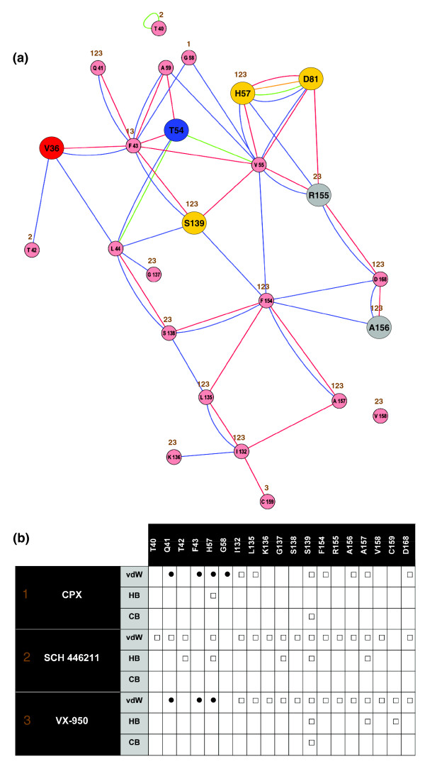
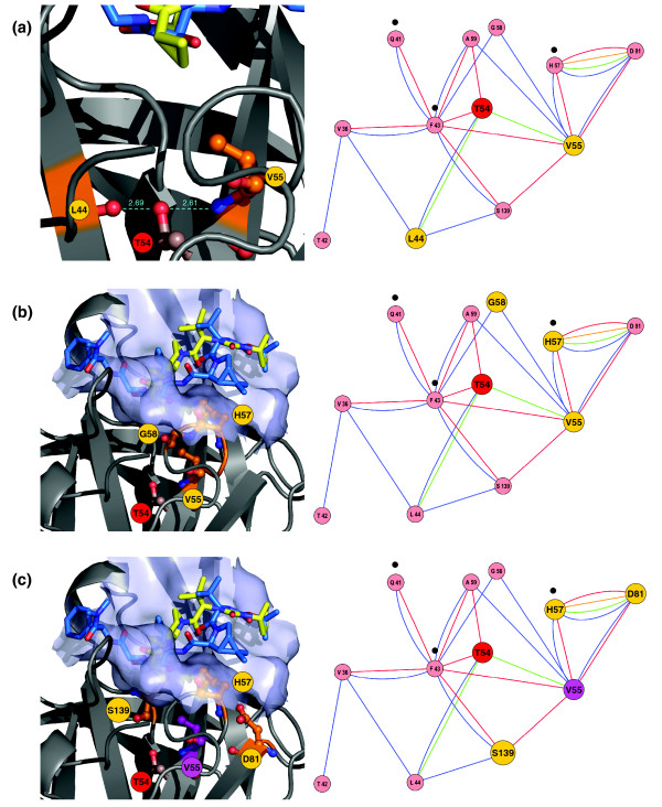
Results: Based on the available experimental structures of NS3-4A, we analyze the binding mode of different ligands. We also investigate the binding mode of VX-950 by protein-ligand docking. A network of non-covalent interactions between amino acids of the protease structure and the interacting ligands is analyzed to discover possible mechanisms of drug resistance. We describe the potential impact of V36 and T54 mutants on the side chain and backbone conformations and on the non-covalent residue interactions. We propose possible explanations for their effects on the antiviral efficacy of drugs and viral fitness. Molecular dynamics simulations of T54A/S mutants and rotamer analysis of V36A/G/L/M side chains support our interpretations. Experimental data using an HCV V36G replicon assay corroborate our findings.
Conclusion: T54 mutants are expected to interfere with the catalytic triad and with the ligand binding site of the protease. Thus, the T54 mutants are assumed to affect the viral replication efficacy to a larger degree than V36 mutants. Mutations at V36 and/or T54 result in impaired interaction of the protease residues with the VX-950 cyclopropyl group, which explains the development of viral breakthrough variants.
Figures

Similar articles
-
Pharmacophore-Assisted Covalent Docking Identifies a Potential Covalent Inhibitor for Drug-Resistant Genotype 3 Variants of Hepatitis C Viral NS3/4A Serine Protease.Viruses. 2024 Aug 3;16(8):1250. doi: 10.3390/v16081250. Viruses. 2024. PMID: 39205224 Free PMC article.
-
Peptidomimetic escape mechanisms arise via genetic diversity in the ligand-binding site of the hepatitis C virus NS3/4A serine protease.Gastroenterology. 2012 Mar;142(3):654-63. doi: 10.1053/j.gastro.2011.11.035. Epub 2011 Dec 7. Gastroenterology. 2012. PMID: 22155364 Free PMC article.
-
Phenotypic characterization of resistant Val36 variants of hepatitis C virus NS3-4A serine protease.Antimicrob Agents Chemother. 2008 Jan;52(1):110-20. doi: 10.1128/AAC.00863-07. Epub 2007 Oct 15. Antimicrob Agents Chemother. 2008. PMID: 17938182 Free PMC article. Clinical Trial.
-
Discovery and development of VX-950, a novel, covalent, and reversible inhibitor of hepatitis C virus NS3.4A serine protease.Infect Disord Drug Targets. 2006 Mar;6(1):3-16. doi: 10.2174/187152606776056706. Infect Disord Drug Targets. 2006. PMID: 16787300 Review.
-
[Telaprevir resistance].Enferm Infecc Microbiol Clin. 2013 Jul;31 Suppl 3:26-32. doi: 10.1016/S0213-005X(13)70121-6. Enferm Infecc Microbiol Clin. 2013. PMID: 24063900 Review. Spanish.
Cited by
-
Pharmacodynamic analysis of a serine protease inhibitor, MK-4519, against hepatitis C virus using a novel in vitro pharmacodynamic system.Antimicrob Agents Chemother. 2012 Mar;56(3):1170-81. doi: 10.1128/AAC.05383-11. Epub 2011 Dec 12. Antimicrob Agents Chemother. 2012. PMID: 22155837 Free PMC article.
-
Resistance-associated variants in chronic hepatitis C patients treated with protease inhibitors.Curr Gastroenterol Rep. 2012 Feb;14(1):47-54. doi: 10.1007/s11894-011-0237-1. Curr Gastroenterol Rep. 2012. PMID: 22161022 Review.
-
NBCZone: Universal three-dimensional construction of eleven amino acids near the catalytic nucleophile and base in the superfamily of (chymo)trypsin-like serine fold proteases.Int J Biol Macromol. 2020 Jun 15;153:399-411. doi: 10.1016/j.ijbiomac.2020.03.025. Epub 2020 Mar 6. Int J Biol Macromol. 2020. PMID: 32151723 Free PMC article.
-
Resistance Mechanisms in Hepatitis C Virus: implications for Direct-Acting Antiviral Use.Drugs. 2017 Jul;77(10):1043-1055. doi: 10.1007/s40265-017-0753-x. Drugs. 2017. PMID: 28497432 Review.
-
Near-Neighbor Interactions in the NS3-4A Protease of HCV Impact Replicative Fitness of Drug-Resistant Viral Variants.J Mol Biol. 2019 May 31;431(12):2354-2368. doi: 10.1016/j.jmb.2019.04.034. Epub 2019 Apr 30. J Mol Biol. 2019. PMID: 31051172 Free PMC article.
References
-
- Hadziyannis SJ, Sette H Jr, Morgan TR, Balan V, Diago M, Marcellin P, Ramadori G, Bodenheimer H Jr, Bernstein D, Rizzetto M, Zeuzem S, Pockros PJ, Lin A, Ackrill AM. PEGASYS International Study Group. Peginterferon-alpha2a and ribavirin combination therapy in chronic hepatitis C: a randomized study of treatment duration and ribavirin dose. Ann Intern Med. 2004;140:346–355. - PubMed
-
- Manns MP, McHutchison JG, Gordon SC, Rustgi VK, Shiffman M, Reindollar R, Goodman ZD, Koury K, Ling M, Albrecht JK. Peginterferon alfa-2b plus ribavirin compared with interferon alfa-2b plus ribavirin for initial treatment of chronic hepatitis C: a randomised trial. Lancet. 2001;358:958–965. doi: 10.1016/S0140-6736(01)06102-5. - DOI - PubMed
-
- Bogen SL, Arasappan A, Bennett F, Chen K, Jao E, Liu YT, Lovey RG, Venkatraman S, Pan W, Parekh T, Pike RE, Ruan S, Liu R, Baroudy B, Agrawal S, Chase R, Ingravallo P, Pichardo J, Prongay A, Brisson J-M, Hsieh TY, Cheng K-C, Kemp SJ, Levy OE, Lim-Wilby M, Tamura SY, Saksena AK, Girijavallabhan V, Njoroge FG. Discovery of SCH446211 (SCH6): a new ketoamide inhibitor of the HCV NS3 serine protease and HCV subgenomic RNA replication. J Med Chem. 2006;49:2750–2757. doi: 10.1021/jm060077j. - DOI - PubMed
Publication types
MeSH terms
Substances
LinkOut - more resources
Full Text Sources
