. 2008 Jan 23:8:6.
doi: 10.1186/1471-213X-8-6.

Lactation failure in Src knockout mice is due to impaired secretory activation

Affiliations

Lactation failure in Src knockout mice is due to impaired secretory activation

Harriet Watkin et al. BMC Dev Biol. .

Abstract

Background: Mammary gland development culminates in lactation and is orchestrated by numerous stimuli and signaling pathways. The Src family of nonreceptor tyrosine kinases plays a pivotal role in cell signaling. In order to determine if Src plays a role in mammary gland development we have examined mammary gland development and function during pregnancy and lactation in mice in which expression of Src has been eliminated.

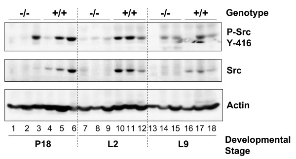
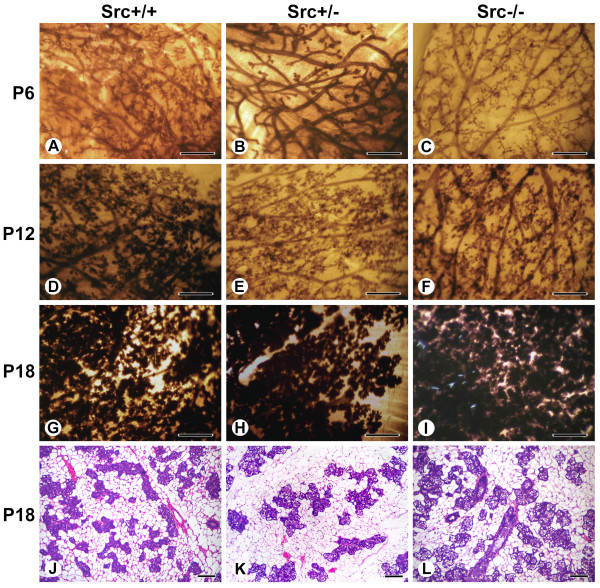
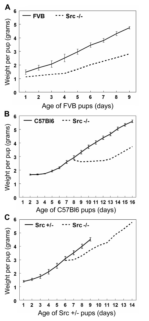
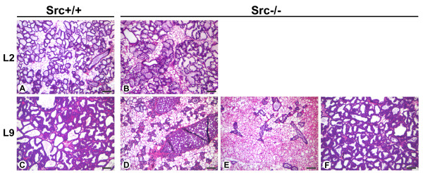
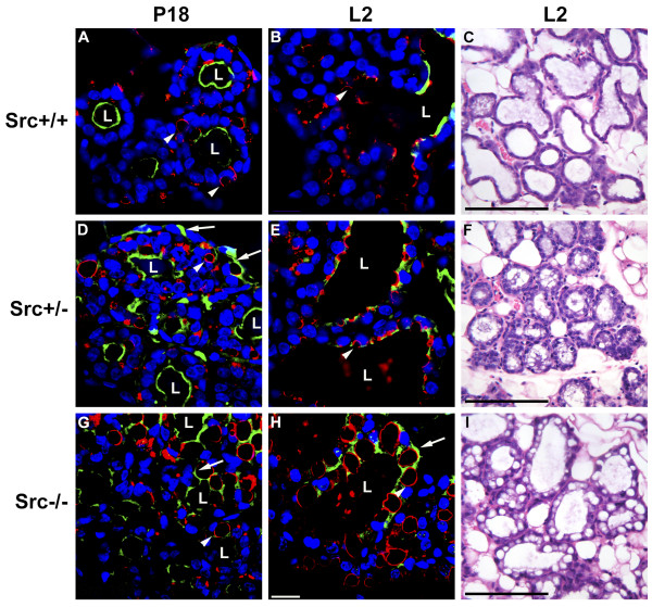
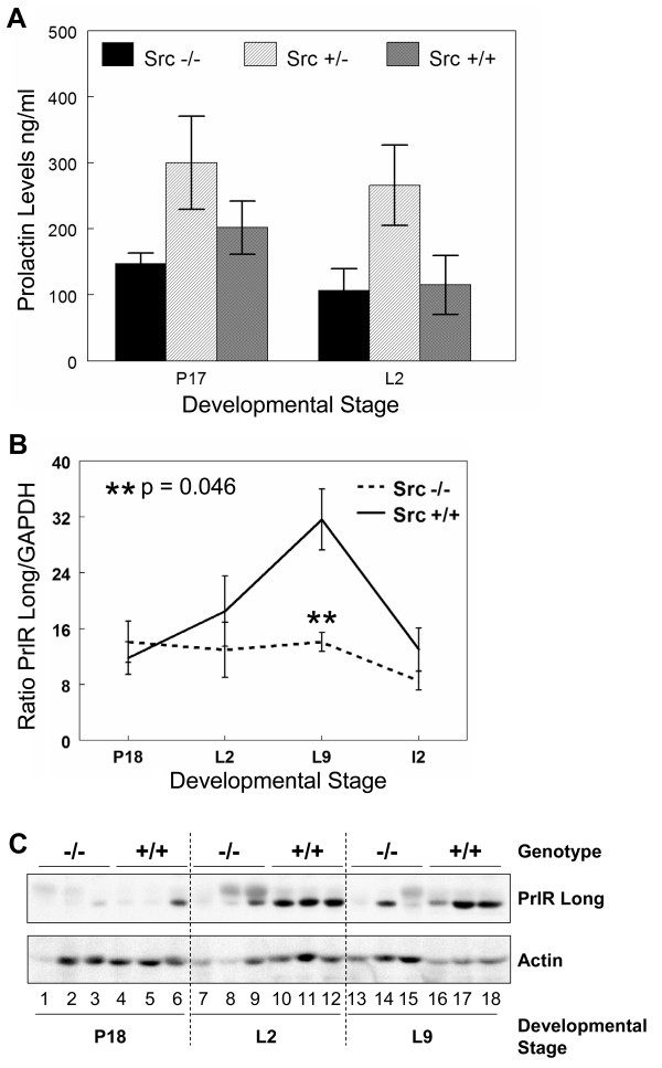
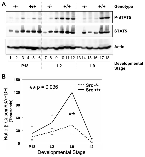
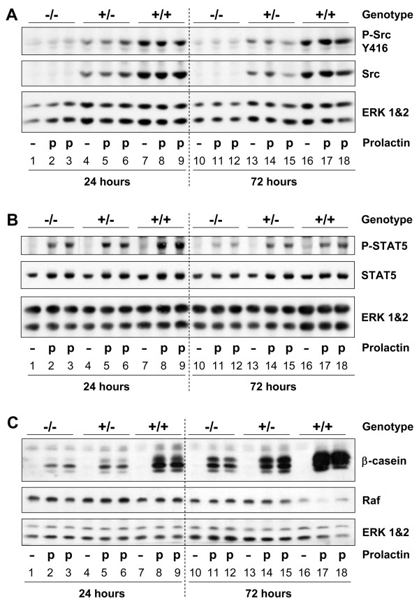
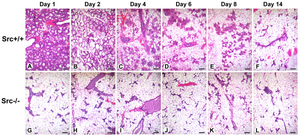
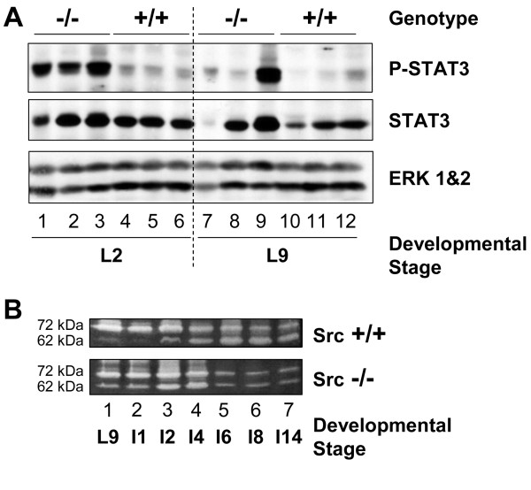
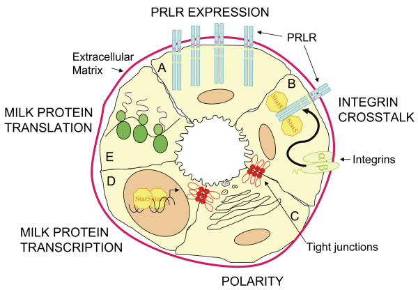
Results: We have characterized a lactation defect in the Src-/- mice which results in the death of over 80% of the litters nursed by Src-/- dams. Mammary gland development during pregnancy appears normal in these mice; however secretory activation does not seem to occur. Serum prolactin levels are normal in Src-/- mice compared to wildtype controls. Expression of the prolactin receptor at both the RNA and protein level was decreased in Src-/- mice following the transition from pregnancy to lactation, as was phosphorylation of STAT5 and expression of milk protein genes. These results suggest that secretory activation, which occurs following parturition, does not occur completely in Src-/- mice. Failed secretory activation results in precocious involution in the mammary glands of Src-/- even when pups were suckling. Involution was accelerated following pup withdrawal perhaps as a result of incomplete secretory activation. In vitro differentiation of mammary epithelial cells from Src-/- mice resulted in diminished production of milk proteins compared to the amount of milk proteins produced by Src+/+ cells, indicating a direct role for Src in regulating the transcription/translation of milk protein genes in mammary epithelial cells.

Conclusion: Src is an essential signaling modulator in mammary gland development as Src-/- mice exhibit a block in secretory activation that results in lactation failure and precocious involution. Src appears to be required for increased expression of the prolactin receptor and successful downstream signaling, and alveolar cell organization.


Figures

Figure 1
Figure 1
Src is expressed and activated in wildtype mammary glands during pregnancy and lactation. The number 4 mammary gland was removed from Src-/- and Src+/+ mice on day 18 of pregnancy (P18) (lanes 1–6), lactation day 2 (L2) (lanes 7–12), and lactation day 9 (L9) (lanes 13–18). Three separate mice were used per genotype and developmental stage. Protein lysates were prepared as described in the Materials and Methods sections, and immunoblotting conducted to detect phosphorylated Src (tyrosine416) (top panel), total Src (middle panel), total actin, as a loading control (bottom panel).
Figure 2
Figure 2
Src deficient mice have normal pregnancy-induced development. Whole mounts of the number 2 and 3 mammary gland of Src+/+ (A, D, G, and J), Src+/- (B, E, H and K) and Src-/- (C, F, I and L) mice at P6 (A-C), P12 (D-F) and P18 (G-I). Histological sections from mammary glands at P18 are also shown for each genotype (J-L). In panels A to I scale bars represent 5 mm. In panels J to L scale bars represent 100 μm.
Figure 3
Figure 3
Pups suckled by Src knockout mice have dramatically reduced weight gain. Graphs demonstrating the weight gain of pups foster nursed by Src-/- mice compared to the weight gain of pups nursed by control dams. Litters were normalized to eight pups. N = 3 for the control mice and error bars depict the s.e.m. in the growth of these control litters. The start of each graph represents day 1 of lactation (first day post-partum) for the nursing mother. The weights of pups nursed by Src-/- mice were recorded for 9 days. A) Weight gain of a single litter of eight newborn FVB pups fostered by Src-/- mouse in comparison to three litters of FVB pups nursed by FVB dams. B) Weight gain of a single litter of eight C57Bl6 pups that were 8 days old when they were cross-fostered onto a Src-/- mouse that was one day post-partum. The growth rate is compared to three litters of C57Bl6 pups nursed by control C57Bl6 dams. C) Weight gain by a single litter of 6-day old Src+/- pups cross-fostered onto a Src-/- mouse that was one day post-partum in comparison to three litters nursed by Src+/- dams.
Figure 4
Figure 4
Altered mammary gland histology during lactation suggests lactation failure in Src-/- mice. Histological sections from the mammary glands of Src+/+ (A and C) and Src-/- (B and D-F) at L2 (A-B), and L9 (C-F). Three sections of mammary glands from Src-/-mice are shown at L9 (D-F). Scale bars represent 100 μm.
Figure 5
Figure 5
Secretory activation does not occur in Src-/- mice following parturition. Histological sections were prepared from the mammary glands of Src+/+ (A-C), Src+/- (D-F), and Src-/- (G-I) mice at P18 (A, D, and G) and L2 (B, C, E, F, H and I). The sections shown in A, B, D, E, G and H were stained anti-ADRP antibody to outline the cytoplasmic lipid droplets (red), Oregon-Green 488-conjugated WPA to outline the surface of secretory alveoli (green), and DAPI to stain the nuclei of mammary epithelial cells (blue). Hematoxylin and eosin-stained sections of mammary glands at L2 are shown in C, F, and I. The bar in panel H represents 10 μm and the bars in panels C, F and I represent 100 μm. Luminal space is indicated by the letter "L", the white arrowheads indicate cytoplasmic lipid droplets, and the white arrows indicates regions where staining with WGA represents atypical patterns not observed in wildtype mice. The black arrows in panel I indicate large cytoplasmic lipid droplets not observed in wildtype mice.
Figure 6
Figure 6
Serum prolactin levels are normal in Src knockout mice however, prolactin receptor expression is reduced. A) Blood was drawn from Src-/-, Src+/- and Src+/+ mice at P17 and L2. Prolactin levels were measured using a radioimmune assay and the amount of PRL plotted as the mean (± SEM) of 5 mice for each genotype at the indicated developmental stages. Welch's t test was used to evaluate the statistical significance (defined as P < 0.05). Src-/- to Src+/+ at P17 P = 0.26, Src+/- to Src+/+ at P17 P = 0.27. Src-/- to Src+/+ at L2 P = 0.89, Src+/- to Src+/+ at L2 P = 0.08. B) Total RNA was isolated from the number 4 mammary gland of wildtype and knockout mice at P18, L2, L9, and I 2; three mice were used per genotype and developmental stage. cDNA was synthesized from 1 μg of total RNA and quantitative RT-PCR was performed using primers and probe specific for the long isoform of the prolactin receptor. PRLR message levels were normalized to GAPDH for each sample and the graph represents the mean (± SEM) relative amount of the triplicate tissue samples. C) The number 4 mammary gland was removed from Src-/- and Src+/+ mice at P18 (lanes 1–6), L 2 (lanes 7–12) and L 9 (lanes 13–18). Three separate mice were used per genotype and developmental stage. Protein lysates were prepared as described in the Materials and Methods sections, and immunoblotting conducted to detect the total amount of PRLR expression (top panel), and the amount of actin, as a loading control (bottom panel).
Figure 7
Figure 7
Downstream signaling from the prolactin receptor is diminished in Src-/- mice. A) The number 4 mammary gland was removed from Src-/- and Src+/+ mice on day 18 of pregnancy (lanes 1–6), lactation day 2 (lanes 7–12) and lactation day 9 (lanes 13–18). Three separate mice were used per genotype and developmental stage. Protein lysates were prepared as described in the Materials and Methods sections, and immunoblotting conducted to detect phosphorylation of STAT5 (top panel), the amount of total STAT5 (middle panel), and the amount of actin, as a loading control (bottom panel). B) Total RNA was isolated from the number 4 mammary gland of wildtype and knockout mice on day 18 of pregnancy, lactation days 2 and 9 and involution day 2, three mice per genotype and developmental stage were used. cDNA was synthesized from 1 μg of total RNA and quantitative RT-PCR was performed using primers and probe specific for β-casein. β-casein message levels were normalized to GAPDH for each sample and the graph represents the mean (± SEM) relative amount of the triplicate tissue samples.
Figure 8
Figure 8
Primary mammary epithelial cell cultures confirm that Src is required for maximal prolactin signaling. Primary mammary epithelial cells isolated from Src-/- (lanes 1–3 and 10–12), Src+/- (lanes 4–6 and 13–15) and Src+/+ (lanes 7–9 and 16–18) mice were cultured on Matrigel in growth medium for 3 days then the medium was changed to differentiation medium and the cells were cultured for a further 24 or 72 hours in the presence or absence of prolactin. Cellular proteins were extracted by lysis in NET buffer. A) Immunoblotting was performed to detect phosphorylation of Src (tyrosine416) (top panel), total levels of Src protein (middle panel) and the amount of ERK 1&2 (bottom panel) as a loading control. B) Immunoblotting was performed to detect phosphorylation of STAT5 (tyrosine694) (top panel), total levels of STAT5 (middle panel) and the amount of ERK 1&2 (bottom panel) as a loading control. C) Immunoblotting was performed to detect β-casein expression (top panel), the levels of Raf and the amount of ERK 1&2 as loading controls (middle and bottom panels).
Figure 9
Figure 9
Involution is accelerated in mammary glands from Src-/- mice. Histological sections from the mammary glands of Src+/+ (A-F) and Src-/- (G-L) mice are shown at I 1 (A, G), I2 (B, H), I4 (C, I), I6 (D, J), I8 (E, K) and I14 (F, L). Scale bars represent 250 μm.
Figure 10
Figure 10
Precocious activation of STAT3 and activation of MMP2 suggest early onset of involution. A) The number 4 mammary gland was removed from Src-/- and Src+/+ mice at P18 (lanes 1–6), L2 (lanes 7–12) and L9 (lanes 13–18). Three separate mice were used per genotype and developmental stage. Protein lysates were prepared as described in the Materials and Methods sections, and immunoblotting conducted to detect phosphorylated STAT3 (tyrosine7705) (top panel), the total amount of STAT3 expression (middle panel), and the amount of ERK 1&2 as a loading control (bottom panel). B) Mammary gland lysates were prepared from Src+/+ (top panel) and Src-/- (bottom panel) mice at L9. I1, I2, I4, I6, I8, and I14. The positions of the 72 kDa molecular weight proenzyme and 62 kDa molecular weight active enzyme are indicated on the left side of the panels.
Figure 11
Figure 11
Model of the pathways required for secretory activation and lactation that are perturbed in Src-/- mice. A) PRLR EXPRESSION. In normal mice PRLR expression increases at parturition, this is absent in the Src-/- mice and they have decreased levels of both PRLR message and protein. B) INTEGRIN CROSSTALK. Integrin-mediated adhesion is essential for STAT5 activation downstream of PRLR signaling and Src may be a required mediator for the β 1 integrin-induced crosstalk. The Src-/- mice have diminished levels of STAT5 phosphorylation. C) POLARITY. Epithelial cell polarity is crucial for secretory activation. The mammary glands of Src-/- mice do not have organized lobuloalveolar structures and many of the epithelial cells do not show restricted apical staining suggesting that Src is required for epithelial cell polarity and organization. D) MILK PROTEIN TRANSCRIPTION. Src-/- mice have significantly less β-casein message compared to wildtype mice. This may be directly related to the decreased levels of STAT5 activation in these cells or Src may also be a required signaling molecule in the additional pathways necessary for milk protein transcription. E) MILK PROTEIN TRANSLATION. Milk protein expression is also regulated at the translational level. Src-/- mice have decreased levels of milk proteins and a possible role for Src in the post-transcriptional regulation has not been investigated.

Similar articles

Cited by

References

    1. Sternlicht MD. The cues that regulate ductal branching and morphogenesis. Breast Cancer Res. 2006;8:201–212. doi: 10.1186/bcr1368. - DOI - PMC - PubMed
    1. Korach KS. Insights from the study of animals lacking functional estrogen receptor. Science. 1994;266:1524–1527. doi: 10.1126/science.7985022. - DOI - PubMed
    1. Korach KS, Couse JF, Curtis SW, Washburn TF, Lindzey J, et al. Estrogen receptor gene disruption: molecular characterization and experimental and clinical phenotypes. Recent Prog Horm Res. 1996;51:159–186. - PubMed
    1. Oakes SR, Rogers RL, Naylor MJ, Ormandy CJ. Prolactin Regulation of Mammary Gland Development. J Mammary Gland Biol Neoplasia. 2008 - PubMed
    1. Humphreys RC, Lydon JP, O'Malley BW, Rosen J. Mammary gland development is mediated by both stromal and epithelial progesterone receptors. Mol Endo. 1997;11:801–811. doi: 10.1210/me.11.6.801. - DOI - PubMed

Publication types

MeSH terms