. 2008 Jan 23:5:1.
doi: 10.1186/1742-6405-5-1.

Human embryonic stem cell (hES) derived dendritic cells are functionally normal and are susceptible to HIV-1 infection

Affiliations

Human embryonic stem cell (hES) derived dendritic cells are functionally normal and are susceptible to HIV-1 infection

Sriram Bandi et al. AIDS Res Ther. .

Abstract

Background: Human embryonic stem (hES) cells hold considerable promise for cell replacement and gene therapies. Their remarkable properties of pluripotency, self-renewal, and tractability for genetic modification potentially allows for the production of sizeable quantities of therapeutic cells of the hematopoietic lineage. Dendritic cells (DC) arise from CD34+ hematopoietic progenitor cells (HPCs) and are important in many innate and adaptive immune functions. With respect to HIV-1 infection, DCs play an important role in the efficient capture and transfer of the virus to susceptible cells. With an aim of generating DCs from a renewable source for HIV-1 studies, here we evaluated the capacity of hES cell derived CD34+ cells to give rise to DCs which can support HIV-1 infection.

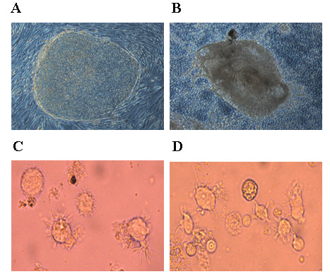
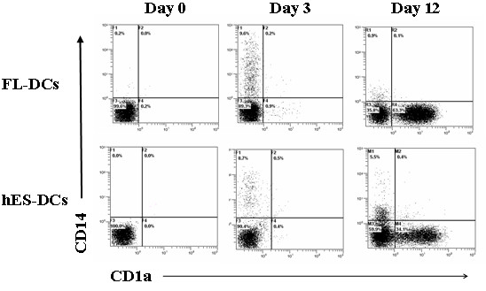
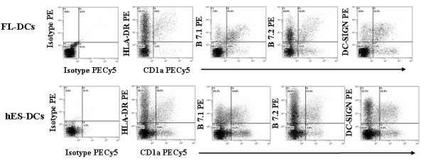
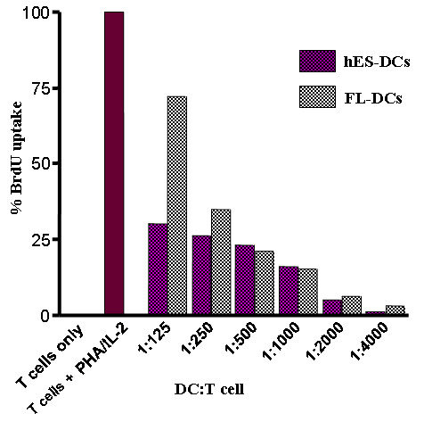
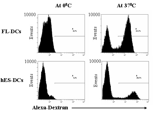
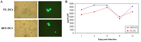
Results: Undifferentiated hES cells were cultured on S17 mouse bone marrow stromal cell layers to derive CD34+ HPCs which were subsequently grown in specific cytokine differentiation media to promote the development of DCs. The hES derived DCs (hES-DC) were subjected to phenotypic and functional analyses and compared with DCs derived from fetal liver CD34+ HPC (FL-DC). The mature hES-DCs displayed typical DC morphology consisting of veiled stellate cells. The hES-DCs also displayed characteristic phenotypic surface markers CD1a, HLA-DR, B7.1, B7.2, and DC-SIGN. The hES-DCs were found to be capable of antigen uptake and stimulating naïve allogeneic CD4+ T cells in a mixed leukocyte reaction assay. Furthermore, the hES-DCs supported productive HIV-1 viral infection akin to standard DCs.

Conclusion: Phenotypically normal and functionally competent DCs that support HIV-1 infection can be derived from hES cells. hES-DCs can now be exploited in applied immunology and HIV-1 infection studies. Using gene therapy approaches, it is now possible to generate HIV-1 resistant DCs from anti-HIV gene transduced hES-CD34+ hematopoietic progenitor cells.


Figures

Figure 1
Figure 1
Derivation of DCs from hES cells: Undifferentiated hES cells were cocultured with S17 mouse stromal cells to derive cystic bodies. Later, purified CD34+ cells derived from cystic bodies and fetal liver were cultured in cytokine media to derive DCs as described in Methods. A and B, representative hES colony and an cystic body respectively. C and D, morphology of DCs differentiated from hES and FL derived CD34+ cells.
Figure 2
Figure 2
FACS analysis of differentiating DCs from hES and FL CD34+ cells: CD34+ cells were cultured in cytokine media and analyzed by FACS for CD14 and CD1a markers at different days by staining with CD1a-PECY5 and CD14-PE conjugated antibodies. Dot plots are representative of triplicate experiments.
Figure 3
Figure 3
Phenotypic analysis of hES-DCs and FL-DCs: hES-DCs and FL-DCs were stained with antibodies CD1a-PECY5, HLA-DR-PE, B7.1-PE, B7.2-PE, and DC-SIGN-PE. Expression of these respective markers was analyzed by FACS. Percent positive cells are indicated in respective plots for each of the cell surface markers. The isotype controls are shown in the left panel. Data is representative of triplicate experiments.
Figure 4
Figure 4
Allogeneic T-cell stimulation by hES and FL derived DCs: The allogeneic stimulatory properties of DCs were assessed in a mixed leukocyte reaction assay using allogeneic T-cells. Graded numbers of sorted and irradiated DCs were co-cultured with 5 × 10⁵ allogeneic T cells. BrdU incorporation was determined by FACS using a PE-conjugated antibody against BrdU. Histograms depict relative percent of BrdU uptake when compared to positive control cells stimulated with IL-2 and PHA. The X-axis is expressed as ratio of stimulator DCs cells to allogeneic responder T cells.
Figure 5
Figure 5
Antigen uptake by hES-DCs: Cultured hES and FL DCs were sorted based on CD1a marker. The cells were then incubated with Alexa-Dextran at 0°C and 37°C for 1 hr and analyzed by FACS as described in Methods. The percent antigen uptake was measured as the difference in percentages between the test (37°C) and control (0°C). The percent positive cells are indicated in the plots for both hES-DCs and FL-DCs. Data are representative of triplicate experiments.
Figure 6
Figure 6
HIV-1 infection of hES and FL DCs: To determine virus susceptibility, FL and hES DCs were infected with a replication competent X4-tropic HIV-GFP reporter virus strain at an m.o.i. of 0.2. At 6 days post infection, cells were visualized by fluorescence microscopy to determine GFP expression in productively infected cells at the single cell level. Phase contrast and fluorescence images are shown for the respective cell types (A). Infected culture supernatants were assayed for viral p24 antigen by ELISA at different days post-infection (B). Data is representative of duplicate experiments.

Similar articles

Cited by

References

    1. Thomson JA, Itskovitz-Eldor J, Shapiro SS, Waknitz MA, Swiergiel JJ, Marshall VS, Jones JM. Embryonic stem cell lines derived from human blastocysts. Science. 1998;282:1145–1147. doi: 10.1126/science.282.5391.1145. - DOI - PubMed
    1. Reubinoff BE, Pera MF, Fong CY, Trounson A, Bongso A. Embryonic stem cell lines from human blastocysts: somatic differentiation in vitro. Nat Biotechnol. 2000;18:399–404. doi: 10.1038/74447. - DOI - PubMed
    1. Zhang SC, Wernig M, Duncan ID, Brustle O, Thomson JA. In vitro differentiation of transplantable neural precursors from human embryonic stem cells. Nat Biotechnol. 2001;19:1129–1133. doi: 10.1038/nbt1201-1129. - DOI - PubMed
    1. Kaufman DS, Hanson ET, Lewis RL, Auerbach R, Thomson JA. Hematopoietic colony-forming cells derived from human embryonic stem cells. Proc Natl Acad Sci USA. 2001;98:10716–10721. doi: 10.1073/pnas.191362598. - DOI - PMC - PubMed
    1. Xu C, Police S, Rao N, Carpenter MK. Characterization and enrichment of cardiomyocytes derived from human embryonic stem cells. Circ Res. 2002;91:501–508. doi: 10.1161/01.RES.0000035254.80718.91. - DOI - PubMed