. 2008 Jan 24;57(2):248-62.
doi: 10.1016/j.neuron.2007.11.027.

Molecular dissociation of the role of PSD-95 in regulating synaptic strength and LTD

Affiliations

Molecular dissociation of the role of PSD-95 in regulating synaptic strength and LTD

Weifeng Xu et al. Neuron. .

Abstract

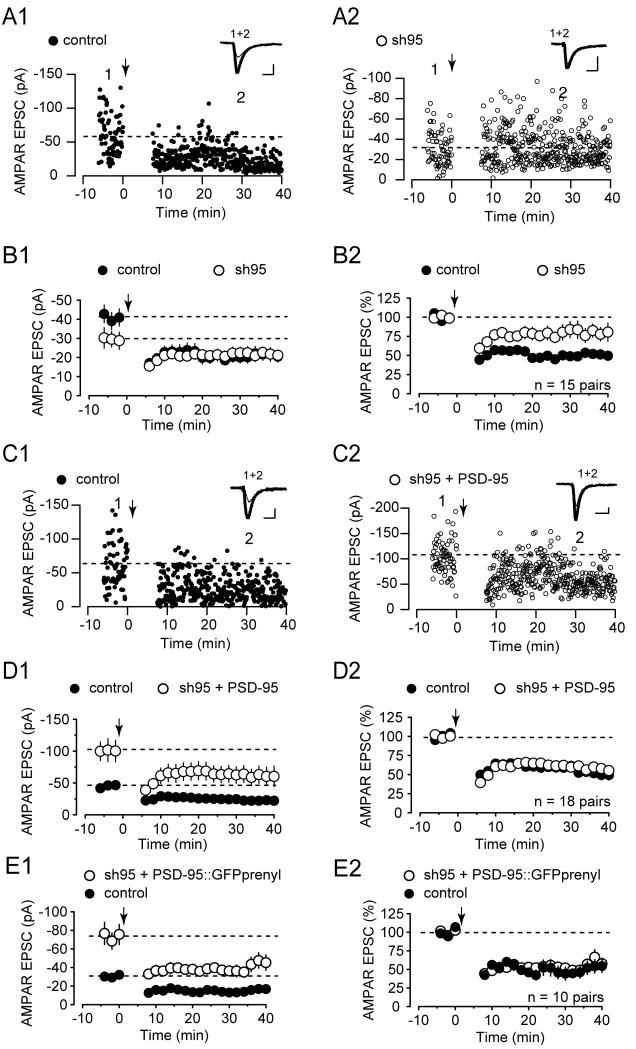
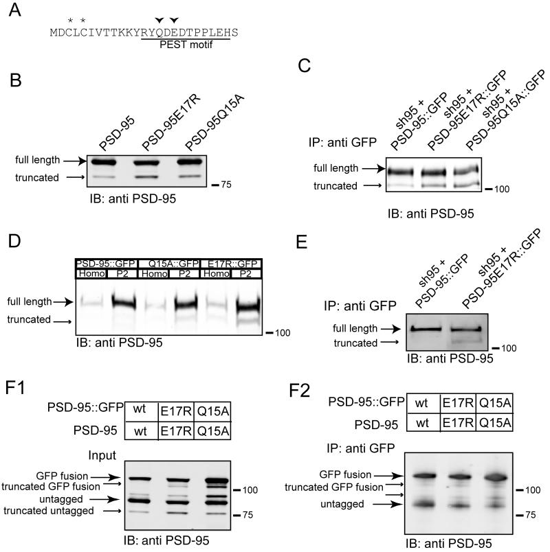
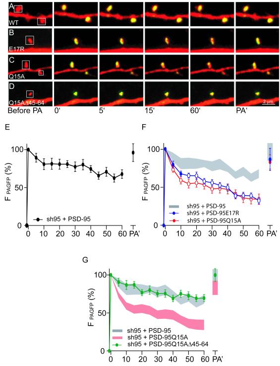
The postsynaptic density protein PSD-95 influences synaptic AMPA receptor (AMPAR) content and may play a critical role in LTD. Here we demonstrate that the effects of PSD-95 on AMPAR-mediated synaptic responses and LTD can be dissociated. Our findings suggest that N-terminal-domain-mediated dimerization is important for PSD-95's effect on basal synaptic AMPAR function, whereas the C-terminal SH(3)-GK domains are also necessary for localizing PSD-95 to synapses. We identify PSD-95 point mutants (Q15A, E17R) that maintain PSD-95's influence on basal AMPAR synaptic responses yet block LTD. These point mutants increase the proteolysis of PSD-95 within its N-terminal domain, resulting in a C-terminal fragment that functions as a dominant negative likely by scavenging critical signaling proteins required for LTD. Thus, the C-terminal portion of PSD-95 serves a dual function. It is required to localize PSD-95 at synapses and as a scaffold for signaling proteins that are required for LTD.


Figures

Figure 1
Figure 1
C-terminal Domains of PSD-95 are Required for Its Effects on Basal AMPAR EPSCs (A) Amplitude of AMPAR EPSCs (left panel, uninfected −34.9 ± 3.5 pA, infected, −59.6 ± 5.7 pA, p < 0.001) and NMDAR EPSCs (middle panel, uninfected 50.7 ± 5.7 pA, infected, 59.1 ± 5.9 pA, p > 0.05) of neurons expressing PSD-95PDZ1+2::GFP are plotted against those of simultaneously recorded uninfected neighboring neurons. (In this and all subsequent panels: gray symbols represent single pairs of recordings; black symbols show mean ± SEM; p values were calculated with a paired Student’s t-test comparing absolute values of paired recordings). Inserts in each panel show sample averaged traces (gray traces, infected neurons; black traces, uninfected neighboring neurons, scale bars, 50 pA/20 ms for AMPAR EPSCs; 50 pA/50 ms for NMDAR EPSCs). Right panels show confocal images of GFP fluorescence (scale bars, 20 μm, left panel, and 2 μm, right panel). (B) Amplitudes of AMPAR EPSCs (uninfected, −60.1 ± 5.5 pA, infected, −30.6 ± 3.8 pA, p < 0.001) and NMDAR EPSCs (uninfected, 64.7 ± 9.7 pA, infected, 49.6 ± 7.4 pA, p < 0.01) of neurons expressing sh95 + PSD-95PDZ1+2::GFP and uninfected neighboring neurons. Right panels show confocal images of GFP fluorescence (scale bars, 20 μm, left panel, and 2 μm, right panel). (C) Amplitudes of AMPAR EPSCs (uninfected, −42.6 ± 4.5 pA, infected, −69.1 ± 5.5 pA, p < 0.001) and NMDAR EPSCs (uninfected, 36.0 ± 5.6 pA, infected, 34.8 ± 3.4 pA, p > 0.05) of neurons expressing PSD-95ΔSH3-GK::GFP and uninfected neighboring neurons. (D) Amplitudes of AMPAR EPSCs (uninfected, −62.0 ± 5.5 pA, infected, −30.3 ± 3.0 pA, p < 0.001) and NMDAR EPSCs (uninfected, 87.8 ± 10.5 pA, infected, 72.0 ± 9.1 pA, p < 0.01) of neurons expressing sh95 + PSD-95ΔSH3-GK-IRES-GFP and uninfected neighboring neurons. (E) Summary (mean ± SEM) of effects of expressing various forms of PSD-95 alone or with sh95 on AMPAR EPSCs calculated as the averaged ratios obtained from pairs of infected and uninfected neighboring neurons (2.17 ± 0.37, 2.35 ± 0.35, 2.35 ± 0.31, 2.72 ± 0.25, 0.51 ± 0.10, 0.56 ± 0.07, 0.59 ± 0.03, respectively, numbers of pairs analyzed are indicated in the bar; n.s. indicates p > 0.05; *** p < 0.001 using an ANOVA Tukey HSD t-test). (F). Summary of effects of same manipulations as in (E) on NMDAR EPSCs (1.21 ± 0.11, 1.36 ± 0.12, 1.23 ± 0.18, 1.20 ± 0.15, 0.80 ± 0.04, 0.85 ± 0.06, 0.88 ± 0.04, respectively, n.s. indicates p > 0.05).
Figure 2
Figure 2
N-terminal Mediated Interactiona are Required for PSD-95 to Affect Basal AMPAR EPSCs (A) Conserved motif in the N-terminus of vertebrate PSD-95. (B) PSD-95 can dimerize through its N-terminus. Western blot of input (top panel) and proteins immnunoprecipitated using GFP antibody (bottom panel) from HEK cells transfected with the indicated constructs and blotted with PSD-95 antibody. (C) Overexpression of PSD-95ΔPEST::GFP does not affect basal AMPAR EPSCs (uninfected, −52.0 ± 6.5 pA, infected, −58.4 ± 7.6 pA, p > 0.05, n=42 pairs) nor NMDAR EPSCs (uninfected, 67.5 ± 7.3 pA, infected, 62.7 ± 6.9 pA, p > 0.05, n=20 pairs) (scale bars, 50 pA/20 ms for AMPAR EPSCs; 50 pA/50 ms for NMDAR EPSCs). (D) Confocal images of GFP from neurons infected with PSD-95ΔPEST::GFP and PSD-95::GFP (scale bars, 20 μm, top panels and 2 μm, bottom panels).
Figure 3
Figure 3
Reduction of LTD by Acute Knockdown of PSD-95 is Rescued by Replacement with Wildtype or Prenylated PSD-95 (A) Sample experiment showing simultaneous recording of LTD of AMPAR EPSCs from an uninfected control cell (A1) and an sh95 infected neighboring cell (A2). Downward arrow in this and all subsequent figures indicates time at which LTD induction protocol was given. In this and all subsequent figures, traces above the graph show averaged EPSCs (20–40 consecutive responses) taken at the times indicated by the numbers on the graph (thick traces, averaged EPSCs from baseline; thin traces, averaged EPSCs after LTD induction; scale bars, 20 pA/20 ms). (B1) Summary graph of LTD of AMPAR EPSCs from pairs of uninfected and sh95 infected cells ( n = 15 pairs). In this and all subsequent summary graphs, points represent mean ± s.e.m. (B2) Summary graph of LTD of AMPAR EPSCs from pairs of uninfected and sh95 infected cells, normalized to the baseline responses of each cell. (n = 15 pairs). (C) Sample experiment illustrating LTD of AMPAR EPSCs from an uninfected control cell (C1) and an sh95+PSD-95::GFP infected neighboring cell (C2) recorded simultaneously. (D1) Summary graph of LTD of AMPAR EPSCs from pairs of uninfected and sh95+PSD-95::GFP infected cells (n = 18 pairs). (D2) Summary graph of LTD of AMPAR EPSCs from pairs of uninfected and sh95+PSD-95::GFP infected cells, normalized to the baseline responses of each cell. (n = 18 pairs). (E1) Summary graph of LTD of AMPAR EPSCs from pairs of uninfected and sh95 + PSD-95::GFPprenyl infected cells ( n = 10 pairs). (E2) Summary graph of LTD of AMPAR EPSCs from pairs of uninfected and sh95 + PSD-95::GFPprenyl infected cells, normalized to the baseline responses of each cell. (n = 10 pairs).
Figure 4
Figure 4
Replacement of Endogenous PSD-95 with E17R or Q15A Point Mutants Rescues Basal AMPAR EPSCs Yet Blocks LTD (A) Amplitude of AMPAR EPSCs of neurons expressing sh95 + PSD-95E17R::GFP (left panel, uninfected −21.7 ±3.0 pA; infected −62.5 ±7.7 pA; p < 0.001) and sh95 + PSD-95Q15A::GFP (middle panel, uninfected −40.4 ±3.8 pA; infected −135.1 ± 14.0 pA; p < 0.001) are plotted against those of simultaneously recorded uninfected neighboring neurons. Right panel shows summary of effects on AMPAR EPSCs of expressing sh95 + PSD-95::GFP, sh95 + PSD-95E17R::GFP, and sh95 + PSD-95Q15A::GFP (2.72 ± 0.25, 3.77 ± 0.66, 3.34 ± 0.34, n.s. indicates p > 0.05). (B) Sample experiment illustrating LTD of AMPAR EPSCs from an uninfected control cell (B1) and an sh95 + PSD-95E17R::GFP infected neighboring cell (B2) recorded simultaneously. (C1) Summary graph of LTD of AMPAR EPSCs from pairs of uninfected and sh95 + PSD-95E17R::GFP infected cells (n = 15 pairs). (C2) Summary graph of LTD of AMPAR EPSCs from pairs of uninfected and sh95 + PSD-95E17R::GFP infected cells, normalized to the baseline responses of each cell. (n = 15 pairs). (D) Sample experiment illustrating LTD of AMPAR EPSCs from an uninfected control cell (D1) and an sh95+PSD-95Q15A::GFP infected neighboring cell (D2) recorded simultaneously. (E1) Summary graph of LTD of AMPAR EPSCs from pairs of uninfected and sh95+PSD-95Q15A::GFP infected cells (n = 12 pairs). (E2) Summary graph of LTD of AMPAR EPSCs from pairs of uninfected and sh95+PSD-95Q15A::GFP infected cells, normalized to the baseline responses of each cell. (n = 12 pairs).
Figure 5
Figure 5
The E17R and Q15A Point Mutants in the N-terminus of PSD-95 Increases Its Truncation (A) Positions of the two point mutations are shown. (B) In HEK293 cells E17R and Q15A mutants show increased amounts of truncated PSD-95 compared to wildtype PSD-95. (C) The increased truncation of E17R and Q15A mutants is present in neurons infected with the indicated replacement viruses. (D) The truncated form of PSD-95 is present in a crude, synaptosomal P2 fraction prepared from cortical neuron cultures infected with the indicated replacement constructs. (E) The increased truncation of the E17R mutant is present in hippocampal slice cultures infected with the indicated replacement viruses. (F) The E17R and Q15A mutants dimerize, but the truncated PSD-95 does not. Western blot of input (F1) and proteins immnunoprecipitated with GFP-antibody (F2) from HEK cells transfected with the indicated constructs.
Figure 6
Figure 6
Over-expression of E17R or Q15A Mutants Blocks LTD (A1) Summary graph of LTD of AMPAR EPSCs from pairs of uninfected and PSD-95E17R::GFP infected cells (n = 17 pairs). (A2) Summary graph of LTD of AMPAR EPSCs from pairs of uninfected and PSD-95E17R::GFP infected cells, normalized to the baseline responses of each cell (n = 17 pairs). (B1) Summary graph of LTD of AMPAR EPSCs from pairs of uninfected and PSD-95Q15A::GFP infected cells (n = 13 pairs). (B2) Summary graph of LTD of AMPAR EPSCs from pairs of uninfected and PSD-95Q15A::GFP infected cells, normalized to the baseline responses of each cell. (n = 13 pairs).
Figure 7
Figure 7
Deletion of Amino Acid Residues 45-64 Prevents the Increased Truncation of the PSD-95Q15A Mutant and Rescues LTD (A) Amino acid sequence of the N-terminal domain of PSD-95Q15AΔ45-64, showing the position of the deletion. (B) Western blot showing that in HEK293 cells PSD-95Q15AΔ45-64 is not truncated. (C) PSD-95Q15AΔ45-64 dimerizes. Western blot of input (upper panel) and proteins immnunoprecipitated with GFP antibody (lower panel) from HEK cells transfected with the indicated constructs. (D) Summary graph of LTD of AMPAR EPSCs from pairs of uninfected and sh95 + PSD-95 Q15AΔ45-64::GFP infected cells (n = 11 pairs). (E) Summary graph of LTD of AMPAR EPSCs from pairs of uninfected and sh95 + PSD-95 Q15AΔ45-64::GFP infected cells, normalized to the baseline responses of each cell (n = 11 pairs).
Figure 8
Figure 8
The E17R and Q15A Point Mutants Show Increased Diffusion Out of Spines Due to Their Truncation (A-D) Sample images of spines from neurons expressing dsRed and the indicated forms of PSD-95 fused to photoactivatable GFP. At time 0′ GFP was photoactivated and changes in fluorescence intensity were monitored at the indicated times. A second photoactivation was performed after 60 min of imaging (PA’). (E) Summary graph of loss of GFP fluorescence from individual spines from cells expressing sh95 and wildtype PSD-95 (n=17/5 spines/cells). (F) Summary graphs of loss of GFP fluorescence from individual spines from cells expressing sh95 with PSD-95E17R (n=25/5 spines/cells) or PSD-95Q15A (n=23/5 spines/cells). Open circles indicate time points at which the values are statistically different (p<0.05) compared to the corresponding timepoint in wildtype cells. For comparison, the summary graph of wildtype PSD-95 (mean ± SEM) is shown in the gray shaded region. (G) Summary graph of loss of GFP fluorescence from individual spines from cells expressing sh95 with PSD-9Q15A5Δ45-64 (n=27/5 spines/cells). For comparison, the summary graphs of wildtype PSD-95 (gray) and Q15A PSD-95 (pink) are also shown.
Figure 9
Figure 9
The Block of LTD by Q15A PSD-95 Requires C-terminal Domain Interactions (A1) Summary graph of LTD of AMPAR EPSCs from pairs of uninfected and PSD-95ΔSH3-GK::GFP infected cells (n = 12 pairs). (A2) Summary graph of LTD of AMPAR EPSCs from pairs of uninfected and PSD-95ΔSH3-GK::GFP infected cells, normalized to the baseline responses of each cell. (n = 12 pairs). (B1) Summary graph of LTD of AMPAR EPSCs from pairs of uninfected and PSD-95Q15AΔSH3-GK::GFP infected cells (n = 11 pairs). (B2) Summary graph of LTD of AMPAR EPSCs from pairs of uninfected and PSD-95Q15AΔSH3-GK::GFP infected cells, normalized to the baseline responses of each cell. (n = 11 pairs). (C1) Summary graph of LTD of AMPAR EPSCs from pairs of uninfected and sh95 + PSD-95L460P::GFP infected cells (n = 10 pairs). (C2) Summary graph of LTD of AMPAR EPSCs from pairs of uninfected and sh95 + PSD-95L460P::GFP infected cells, normalized to the baseline responses of each cell (n = 10 pairs). (D1) Summary graph of LTD of AMPAR EPSCs from uninfected (n=8) and PSD-95Q15AL460P::GFP infected cells (n = 11). Data includes 5 pairs of simultaneously recorded uninfected and infected cells, and additional cells recorded individually from slice cultures injected with lentiviruses expressing PSD-95Q15AL460P::GFP. (D2) Summary graph of LTD of AMPAR EPSCs from uninfected (n=8) and PSD-95Q15AL460P::GFP infected cells (n = 11), normalized to the baseline responses of each cell.
Figure 10
Figure 10
Summary of the Effect of PSD-95 Manipulations on Basal Transmission and LTD (A) Summary of the effects of PSD-95 manipulations on LTD. Percentage of the baseline response at 35-40 mins after LTD induction is plotted. Open bars, infected cells; gray bars, uninfected cells. (B) Left panel: model for the role of PSD-95 in basal synaptic AMPAR function. Left side of spine shows the normal condition with endogenous PSD-95 present. Right side shows the consequences of overexpressing PSD-95PDZ1+2, which can interact with endogenous PSD-95 and recruit additional AMPARs. Replacing endogenous PSD-95 with PSD-95PDZ1+2 or expressing PSD-95C3,5S or PSD-95ΔPEST leaves these proteins outside the spine because they cannot dimerize with endogenous PSD-95. (Whether PSD-95ΔPEST is palmitoylated is not known.) Right panel: model for the role of PSD-95 as a signaling scaffold for LTD. Left side of spine shows the normal condition in which PSD-95 interacts with a complex of signaling proteins that is activated by NMDAR-mediated influx of calcium (red arrow) during LTD induction. Right side shows truncated PSD-95 interacting with the signaling complex away from the NMDAR such that it cannot be activated by calcium.

References

    1. Beique JC, Andrade R. PSD-95 regulates synaptic transmission and plasticity in rat cerebral cortex. J. Physiol. 2003;546:859–867. - PMC - PubMed
    1. Beique JC, Lin DT, Kang MG, Aizawa H, Takamiya K, Huganir RL. Synapse-specific regulation of AMPA receptor function by PSD-95. Proc. Natl. Acad. Sci. U. S. A. 2006;103:19535–19540. - PMC - PubMed
    1. Bloodgood BL, Sabatini BL. Neuronal activity regulates diffusion across the neck of dendritic spines. Science. 2005;310:866–869. - PubMed
    1. Bredt DS, Nicoll RA. AMPA receptor trafficking at excitatory synapses. Neuron. 2003;40:361–379. - PubMed
    1. Carroll RC, Lissin DV, von Zastrow M, Nicoll RA, Malenka RC. Rapid redistribution of glutamate receptors contributes to long-term depression in hippocampal cultures. Nature. Neurosci. 1999;2:454–460. - PubMed

Publication types

MeSH terms

Substances