. 2008 Feb;34(2):166-71.
doi: 10.1016/j.joen.2007.11.021.

Characterization of the apical papilla and its residing stem cells from human immature permanent teeth: a pilot study

Affiliations

Characterization of the apical papilla and its residing stem cells from human immature permanent teeth: a pilot study

Wataru Sonoyama et al. J Endod. 2008 Feb.

Abstract

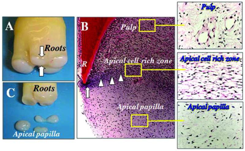
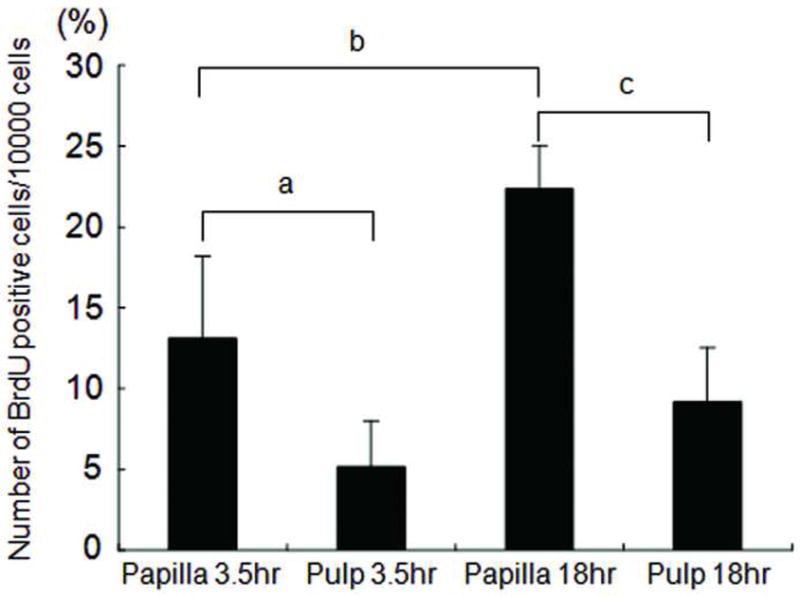
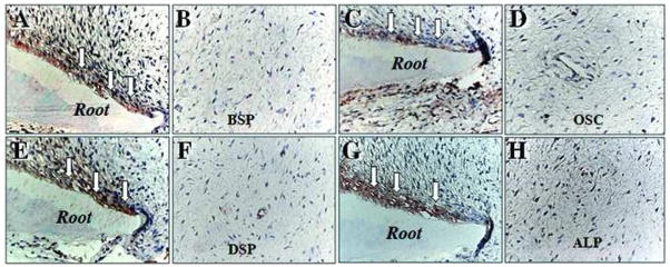
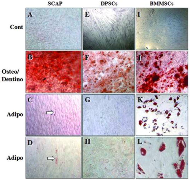
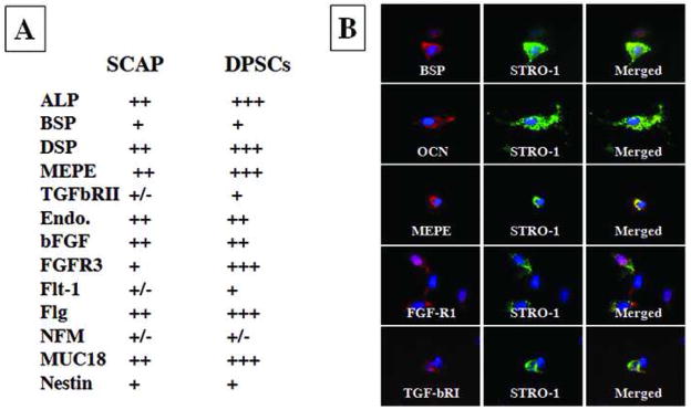
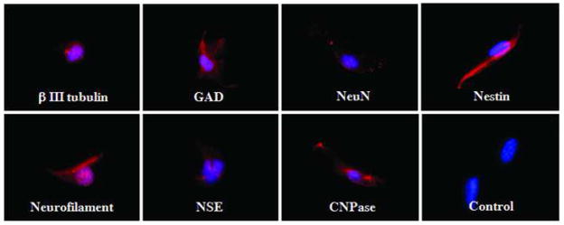
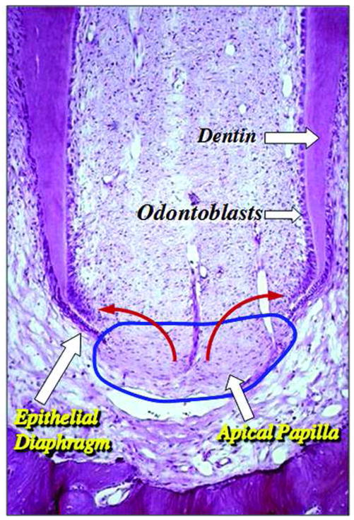
Mesenchymal stem cells (MSCs) have been isolated from the pulp tissue of permanent teeth (dental pulp stem cells or DPSCs) and deciduous teeth (stem cells from human exfoliated deciduous teeth). We recently discovered another type of MSCs in the apical papilla of human immature permanent teeth termed stem cells from the apical papilla (SCAP). Here, we further characterized the apical papilla tissue and stem cell properties of SCAP using histologic, immunohistochemical, and immunocytofluorescent analyses. We found that the apical papilla is distinctive to the pulp in terms of containing less cellular and vascular components than those in the pulp. Cells in the apical papilla proliferated 2- to 3-fold greater than those in the pulp in organ cultures. Both SCAP and DPSCs were as potent in osteo/dentinogenic differentiation as MSCs from bone marrows, whereas they were weaker in adipogenic potential. The immunophenotype of SCAP is similar to that of DPSCs on the osteo/dentinogenic and growth factor receptor gene profiles. Double-staining experiments showed that STRO-1 coexpressed with dentinogenic markers such as bone sialophosphoprotein, osteocalcin, and growth factors FGFR1 and TGFbetaRI in cultured SCAP. Additionally, SCAP express a wide variety of neurogenic markers such as nestin and neurofilament M upon stimulation with a neurogenic medium. We conclude that SCAP are similar to DPSCs but a distinct source of potent dental stem/progenitor cells. Their implications in root development and apexogenesis are discussed.


Figures

Fig. 1
Fig. 1. Anatomy of apical papilla
(A) An extracted human third molar depicting root attached to the root apical papilla (open arrows) at developmental stage. (B) Hematoxylin and eosin (H & E) staining of human developing root (R) depicting epithelial diaphragm (open arrows) and apical cell rich zone (open arrowheads). (C) Harvested root apical papilla for stem cell isolation.
Fig. 2
Fig. 2. Cell proliferation in pulp and apical papilla in cultures
In vitro BrdU labeling after 3.5 and 18 hs. Representative data (out of three independence experiments) indicate the numbers of BrdU positive cells in the pulp compared with those in the apical papilla of the same tooth. Four randomly selected fields per sample were used to count the positively stained cells under the microscope. Error bars: SD; a, p=0.0091; b, p<0.0001; c, p=0.0002.
Fig. 3
Fig. 3. Immunocytofluorescent staining of osteo/dentinogenic markers in pulp and apical papilla
Odontoblasts (open arrows) express osteo/dentinogenic markers including BSP (A), OCN (C), DSP (E), and ALP (G). Apical papilla showed a negative immunohistochemical staining for BSP (B), OSC (D), DSP (F), and ALP (H).
Fig. 4
Fig. 4. Osteo-/dentino- and adipo-genesis of stem cells
SCAP (A–D), DPSCs (E–H) and BMMSCs (I–L). Control unstimulated (A, E, I); osteo/dentinogenic stimulation of cells for 8 weeks (B, F, J); adipogenic stimulation for 3 weeks (C–L). Open arrows indicate oil red O stain. Data are representative from several independent experiments [original magnifications, E, I: 50x; B, F, G, J, K: 100x; A, C, D, H, L: 200x]
Fig. 5
Fig. 5. Immunophenotype of SCAP
(A) SCAP and DPSCs express similar osteo/dentinogenic markers and growth factor receptors. SCAP express less amounts of DSP, MEPE, TGFβRII, FGFR3, Flt-1, Flg, and MUC18. +++, strong; ++, moderate; +, weak; −, negative. (B) Double staining of SCAP showing STRO-1 co-expressed with osteo/dentinogenic markers such as BSP, OCN, MEPE and growth factor receptors FGFR1 and TGFβRI. Cell nuclei exhibit in blue fluorescence by DAPI counter stain. Expression of markers showing either in red of green fluorescence. Images are representative data of independent experiments with consistent results.
Fig. 6
Fig. 6. Expression of neural markers in SCAP
Immunocytofluorescent staining of cultured SCAP expressing a variety of neural markers (red fluorescence) including βIII tubulin, GAD, NeuN, nestin, GFAP, neurofilament M, NSE, and CNPase when cultured in neurogenic medium for 4 weeks. Blue fluorescence is the cell nucleus. Images are representative data of independent experiments with consistent results.
Fig. 7
Fig. 7. Diagram of hypothetical cell source for root dentin development
Apical papilla (blue circle) contains stem/progenitor cells which may be the source of the odontoblasts that produce root dentin (red arrows). [Original histological image of immature tooth from Macacca irus monkey, courtesy of Dr. C. Torneck; original image published in Principles and Practice of Endodontics, 3rd ed., chapter 2, page 6, Fig. 2--2; permission of image reuse obtained from Elsevier Ltd.]

References

    1. Gronthos S, Mankani M, Brahim J, Robey PG, Shi S. Postnatal human dental pulp stem cells (DPSCs) in vitro and in vivo. Proc Natl Acad Sci U S A. 2000;97:13625–30. - PMC - PubMed
    1. Miura M, Gronthos S, Zhao M, et al. SHED: stem cells from human exfoliated deciduous teeth. Proc Natl Acad Sci U S A. 2003;100:5807–12. - PMC - PubMed
    1. Sonoyama W, Liu Y, Fang D, et al. Mesenchymal stem cell-mediated functional tooth regeneration in Swine. PLoS ONE. 2006;1:e79. - PMC - PubMed
    1. D’Souza R. Development of the pulpodentin complex. In: Goodis KMHaHE., editor. Seltzer and Bender’s Dental Pulp. Quintessence Publishing Co, Inc; Carol Stream, IL: 2002.
    1. Sonoyama W, Seo BM, Yamaza T, Shi S. Human Hertwig’s Epithelial Root Sheath Cells Play Crucial Roles in Cementum Formation. J Dent Res. 2007;86:594–9. - PubMed

Publication types

MeSH terms