. 2008 Jan;15(1):5-11.
doi: 10.1016/j.chembiol.2007.11.013.

Analysis of the substrate specificity of the Dim-5 histone lysine methyltransferase using peptide arrays

Affiliations

Analysis of the substrate specificity of the Dim-5 histone lysine methyltransferase using peptide arrays

Philipp Rathert et al. Chem Biol. 2008 Jan.

Abstract

Histone methylation is an epigenetic mark essential for gene regulation and development. We introduce peptide SPOT synthesis to study sequence specificity of the Dim-5 histone-3 lysine-9 methyltransferase. Dim-5 recognizes R8-G12 of the H3 tail with T11 and G12 being the most important specificity determinants. Exchange of H3 tail residue S10 and T11 by E strongly reduced methylation by Dim-5, suggesting that phosphorylation of S10 or T11 may regulate the activity of Dim-5. In the Dim-5/peptide structure, E227 interacts with H3R8 and D209 with H3-S10. Mutations of E227 or D209 caused predictable changes in the substrate preference, illustrating that peptide recognition of histone methyltransferases can be altered by protein design. Comparative analyses of peptide arrays with wild-type and mutant enzymes, therefore, are well suited to investigate the target specificity of protein methyltransferases and study epigenetic crosstalk.


Figures

Figure 1
Figure 1. Application of the Peptide SPOT Synthesis to Study the Activity of Histone Methyltransferases
(A) Example of the methylation of peptides on solid support. After SPOT synthesis of the wild-type histone H3 N-terminal tail sequence (H3) and a K9A variant, the membrane was incubated with Dim-5 and radioactive AdoMet. Methylation was visualized by fluorography. (B and C) The methylation rate of peptide spots increases linearly with enzyme concentration and time. (D) Alanine scan of H3 tail methylation by Dim-5. In this assay, all 21 positions of the H3 tail were exchanged individually against alanine. The spot labeled with wt contains the wild-type H3 tail sequence. (E) Quantitative analysis of four independent alanine scans indicating the average activity and standard error for each target peptide.
Figure 2
Figure 2. Specificity of Peptide Methylation by Dim-5
(A) Example of one full H3 peptide tail array. The sequence of the H3 tail is given on the horizontal axis. Each residue was exchanged against all 20 natural amino acid residues (as indicated on the vertical axis) and the relative efficiency of methylation by Dim-5 analyzed. (B) Compilation of the results of the peptide scan experiments with Dim-5. Data are averaged numbers from three independent experiments. (C) Distribution of standard deviations for the data shown in (B) after normalizing full activity to 1.0. (D) Bar diagram showing the discrimination factors of Dim-5 at the positions tested. (E) Compilation of the specificity of Dim-5 for its target peptide.
Figure 3
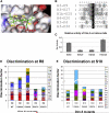
Figure 3. Analysis of Dim-5 Peptide Contacts
(A) Structure of the Dim-5 binding cleft (Zhang et al., 2002). H3 peptide residues from A7-G12 are indicated in green; Dim-5 residues contacting the peptide (D209 and E227) are shown in red. For clarity of visualization, the side chains of D209 and E227 were not included in the calculation of the surface of Dim-5. (B) Alignment of some lysine residues in the different histone tails. (C) Relative rates of modification of H3K9, H3K27, H4K20, and H1bK25 peptides by Dim-5. Error bars denote the standard deviation of the mean. (D) Substrate discrimination of Dim-5 wild-type and mutants on H3 tail substrates with all 20 natural amino acid substitutions at position R8. (E) Substrate discrimination of Dim-5 wild-type and mutants on H3 tail substrates with all 20 natural amino acid substitutions at position S10.

Similar articles

Cited by

References

    1. Berger SL. The complex language of chromatin regulation during transcription. Nature. 2007;447:407–412. - PubMed
    1. Bialek K, Swistowski A, Frank R. Epitope-targeted proteome analysis: towards a large-scale automated protein-protein-interaction mapping utilizing synthetic peptide arrays. Anal. Bioanal. Chem. 2003;376:1006–1013. - PubMed
    1. Cheng X, Collins RE, Zhang X. Structural and sequence motifs of protein (histone) methylation enzymes. Annu. Rev. Biophys. Biomol. Struct. 2005;34:267–294. - PMC - PubMed
    1. Egger G, Liang G, Aparicio A, Jones PA. Epigenetics in human disease and prospects for epigenetic therapy. Nature. 2004;429:457–463. - PubMed
    1. Feinberg AP, Tycko B. The history of cancer epigenetics. Nat. Rev. Cancer. 2004;4:143–153. - PubMed

Publication types

LinkOut - more resources