. 2008 May 9;283(19):13031-4.
doi: 10.1074/jbc.M800549200. Epub 2008 Jan 23.

Identification of a critical tyrosine residue in caspase 8 that promotes cell migration

Affiliations

Identification of a critical tyrosine residue in caspase 8 that promotes cell migration

Simone Barbero et al. J Biol Chem. .

Abstract

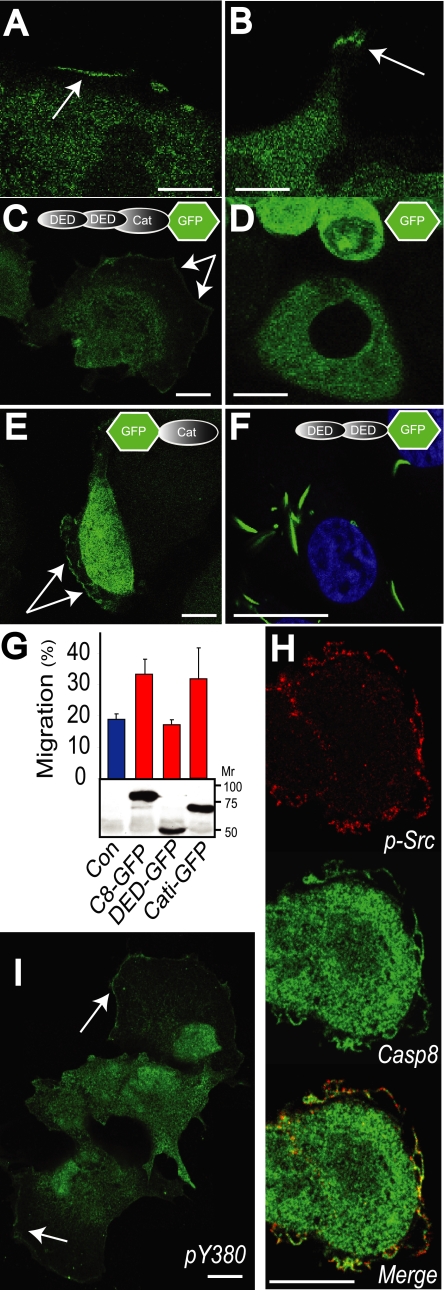
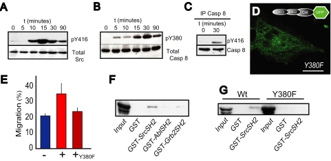
Caspase 8 is a critical upstream initiator of programmed cell death but, paradoxically, has also been shown to promote cell migration. Here, we show that tyrosine 380 in the linker loop of human caspase 8 is a critical switch determining caspase 8 function. Our studies show that, in addition to its cytosolic distribution, caspase 8 is recruited to lamella of migrating cells. Although the catalytic domain of caspase 8 is sufficient for recruitment and promotion of cell migration, catalytic activity per se is not required. Instead, we find that integrin-mediated adhesion promotes caspase 8 phosphorylation on tyrosine 380. Accordingly, mutation of this site compromises localization to the periphery and the potentiation of cell migration. Mechanistically, this linker region of caspase 8 acts as a Src homology 2 binding site. In particular, tyrosine 380 is critical for interaction with Src homology 2 domains. The results identify a novel mechanism by which caspase 8 is recruited to the lamella of a migrating cell, promoting cell migration independent of its protease activity.


Figures

FIGURE 1.
FIGURE 1.
Caspase 8 is recruited to the periphery of migrating and spreading cells. A and B, NB7 cells expressing caspase 8 were allowed to attach to a fibronectin substrate at confluence for 1 h, and the monolayer was wounded and cells allowed to migrate into the wound for a further 2 h. Cells were then fixed and stained for caspase 8 (green signal). To assess the localization of caspase 8 in live cells, caspase 8-GFP fusion proteins were expressed in caspase 8-deficient NB7 cells and imaged (green signal). The localization of full-length GFP-caspase 8 containing both death effector (DED) and catalytic (Cat) domains (C), the GFP protein alone (D), and the GFP-tagged inactive mutant of the catalytic domain C360A (E) are shown in live cells. F, cells expressing GFP-death effector domains were fixed in paraformaldehyde and stained with the nuclear stain TOPRO (blue signal) for contrast. G, the migration of NB7 cells expressing caspase 8 mutants into a wound was assessed after 20 h. Caspase 8-deficient NB7 cells or NB7 cells transiently expressing full-length GFP-caspase 8, GFP-death effector domains, or caspase 8 cells were seeded on a fibronectin substrate at confluence and wounded and the wound allowed to close for 20 h. Data shown are the means ± S.E. from six determinations in a representative assay. To confirm the expression of the caspase 8 constructs in the NB7 cells, lysates were separated via PAGE and immunoblotted with anti-GFP to resolve the GFP tag (lower panel). H, cells expressing the C8-GFP construct were fixed and stained for active Src using antisera to Src pY416 as a reporter (red channel). I, immunofluorescence staining for caspase 8 using pY380-specific antisera was performed in randomly migrating COS7 cells (green signal). The bars shown in each image A–F and H–I are 10 μm.
FIGURE 2.
FIGURE 2.
Phosphorylated caspase 8 associates with Src via the SH2 domain. A, NB7 neuroblastoma cells were allowed to attach to and spread on fibronectin-coated surfaces, lysed, and subjected to immunoblot analysis for active Src using anti-pY416 antisera. B, similarly, NB7 cells stably reconstituted for caspase 8 were allowed to attach to fibronectin-coated surfaces and subjected to immunoblot analysis using anti pY380 antisera to detect phospho-caspase 8. C, cells were allowed to attach and spread on fibronectin substrate for 30 min and then lysed in radioimmune precipitation buffer and the complexes subjected to immunoprecipitation using monoclonal antibodies specific for caspase 8. Immunoprecipitated complexes were resolved on 10% SDS gels and subjected to immunoblot analysis using rabbit antisera to p416 (active Src) or caspase 8 to confirm the immunoprecipitation. D, NB7 cells were reconstituted with GFP-C8 bearing the Y380F mutant and allowed to attach to fibronectin-coated surfaces, and the localization of C8 was assessed by immunofluorescence microscopy. E, the NB7-Y380F cells were assessed for their capacity to migrate in a wounding assay, as described above. Data shown are the means ± S.E. from six determinations in a representative assay. F, human embryonic kidney cells expressing active Src kinase (Y527F) were transfected with caspase 8. Cells lysates were then subjected to pull down with 2 μg of GST-SH2 domains from Abl, Grb2, or Src to isolate the phospho-caspase 8. G, similarly, human embryonic kidney(Y527F) cells were transfected with either wild-type caspase 8 or the Y380F mutant and then subjected to pulldown analysis with the GST-SrcSH2 fusion protein, resolved on 10% SDS gels, and immunoblotted to detect caspase 8.

References

    1. Muzio, M., Chinnaiyan, A. M., Kischkel, F. C., O'Rourke, K., Shevchenko, A., Ni, J., Scaffidi, C., Bretz, J. D., Zhang, M., Gentz, R., Mann, M., Krammer, P. H., Peter, M. E., and Dixit, V. M. (1996) Cell 85 817–827 - PubMed
    1. Pop, C., Fitzgerald, P., Green, D. R., and Salvesen, G. S. (2007) Biochemistry 46 4398–4407 - PubMed
    1. Juo, P., Kuo, C. J., Yuan, J., and Blenis, J. (1998) Curr. Biol. 8 1001–1008 - PubMed
    1. Varfolomeev, E. E., Schuchmann, M., Luria, V., Chiannilkulchai, N., Beckmann, J. S., Mett, I. L., Rebrikov, D., Brodianski, V. M., Kemper, O. C., Kollet, O., Lapidot, T., Soffer, D., Sobe, T., Avraham, K. B., Goncharov, T., Holtmann, H., Lonai, P., and Wallach, D. (1998) Immunity 9 267–276 - PubMed
    1. Kang, T. B., Ben-Moshe, T., Varfolomeev, E. E., Pewzner-Jung, Y., Yogev, N., Jurewicz, A., Waisman, A., Brenner, O., Haffner, R., Gustafsson, E., Ramakrishnan, P., Lapidot, T., and Wallach, D. (2004) J. Immunol. 173 2976–2984 - PubMed

Publication types