. 2008 Mar 28;283(13):8723-35.
doi: 10.1074/jbc.M706105200. Epub 2008 Jan 23.

Glucagon-like peptide-1 activation of TCF7L2-dependent Wnt signaling enhances pancreatic beta cell proliferation

Affiliations

Glucagon-like peptide-1 activation of TCF7L2-dependent Wnt signaling enhances pancreatic beta cell proliferation

Zhengyu Liu et al. J Biol Chem. .

Abstract

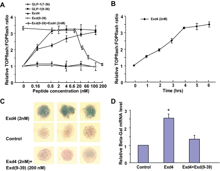
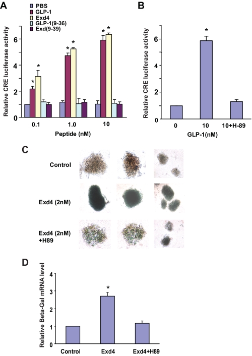
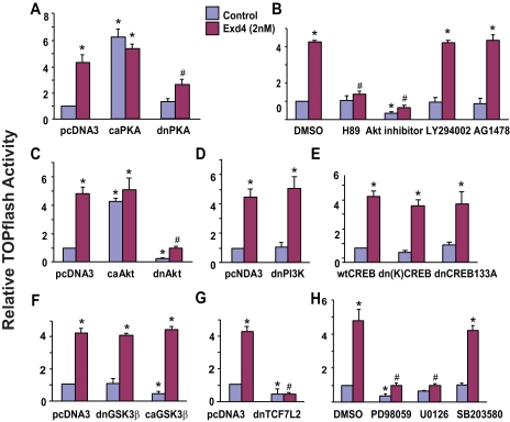
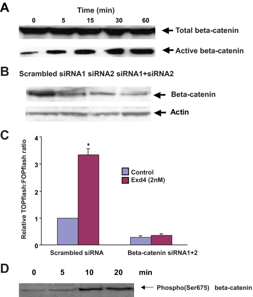
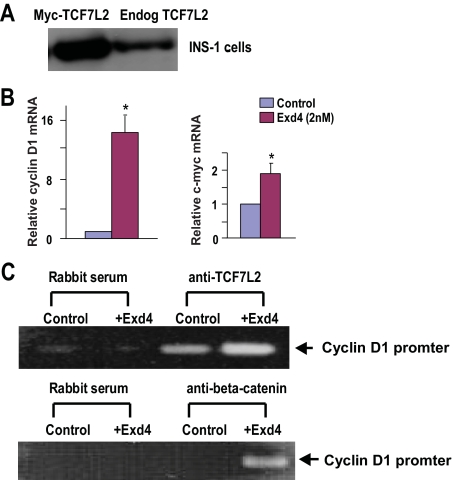
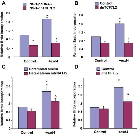
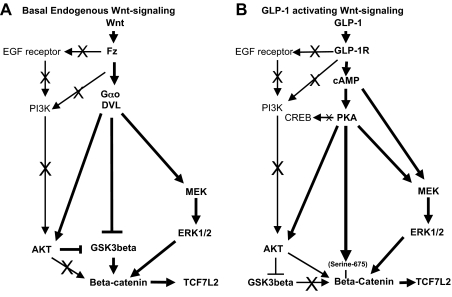
The insulinotropic hormone GLP-1 (glucagon-like peptide-1) is a new therapeutic agent that preserves or restores pancreatic beta cell mass. We report that GLP-1 and its agonist, exendin-4 (Exd4), induce Wnt signaling in pancreatic beta cells, both isolated islets, and in INS-1 cells. Basal and GLP-1 agonist-induced proliferation of beta cells requires active Wnt signaling. Cyclin D1 and c-Myc, determinants of cell proliferation, are up-regulated by Exd4. Basal endogenous Wnt signaling activity depends on Wnt frizzled receptors and the protein kinases Akt and GSK3beta but not cAMP-dependent protein kinase. In contrast, GLP-1 agonists enhance Wnt signaling via GLP-1 receptor-mediated activation of Akt and beta cell independent of GSK3beta. Inhibition of Wnt signaling by small interfering RNAs to beta-catenin or a dominant-negative TCF7L2 decreases both basal and Exd4-induced beta cell proliferation. Wnt signaling appears to mediate GLP-1-induced beta cell proliferation raising possibilities for novel treatments of diabetes.


Figures

FIGURE 1.
FIGURE 1.
Exd4 and GLP-1-(7-36) but not Exd4-(9-39) and GLP-1-(9-36) activate Wnt signaling in INS-1 cells and isolated mouse islets. Wnt signaling is indicated by the value of TOPflash luciferase activity divided by FOPflash activity. A, ratio of TOPflash to FOPflash activity in INS-1 cells treated with increasing doses of GLP-1-(7-36), GLP-1-(9-36), Exd4, and Exd-(9-39). Increasing doses of Exd-(9-39) antagonize Exd4 activation of Wnt signaling. The x axis scale for the Exd4 and Exd-(9-39) experiment refers to the peptide concentration of Exd-(9-39) antagonist added at increasing doses to a fixed concentration (2 nm) of the Exd4 agonist. All values are relative to the zero peptide concentration. Data are normalized for transfection efficiency by co-transfected β-galactosidase and represent means ± S.D. of three experiments. B, a time course of the TOPflash:FOPflash ratio of INS-1 cell treated with 2 nm Exd4. C, phase contrast microscopic images of islets isolated from TOPGAL mice treated with Exd4 (top) or vehicle (middle) or Exd4 and Exd-(9-39) (bottom). D, real time RT-PCR results of lacZ transcription in islets of TOPGAL mice treated with Exd4 or vehicle or Exd4 and Exd-(9-39). Expression values are displayed relative to vehicle-treated condition (control). Data are normalized by using the housekeeping gene GAPDH as internal control. Statistical significance is depicted as * (p < 0.05) when compared with control values.
FIGURE 2.
FIGURE 2.
The GLP-1 receptor is coupled to the cAMP/PKA/CREB signaling pathway in INS-1 cells and isolated mouse islets. GLP-1 and Exd4, but not GLP-1-(9-36) and Exd-(9-39), activate a CRE transcriptional reporter in INS-1 cells. INS-1 cells expressing the pCRE-LUC reporter plasmid were treated for 4 h at 37. A, dose-response of CRE luciferase reporter gene expression in response to increasing concentrations of GLP-1, Exd4, GLP-1-(9-36), and Exd-(9-39). B, stimulation of CRE luciferase gene expression with GLP-1 (10 nm) was abolished by co-incubation of H-89 (10 μm). All values are relative to the 1st (leftmost) bar. Data are normalized for transfection efficiency by co-transfected β-galactosidase and represent means ± S.D. of three experiments. Statistical significance is depicted as * (p < 0.05) when compared with control values. C, mouse islets isolated ex vivo from TOPGAL Wnt signaling reporter mice were incubated for 4 h with Exd4 (2 nm) in the presence and absence of the cAMP/PKA inhibitor H89 (10 μm) (see legend to Fig. 1C). D, real time RT-PCR of lacZ transcription in islets of TOPGAL mice treated with vehicle or Exd4 or Exd4 and H89. Expression values are displayed relative to vehicle-treated control. Data are normalized by using the housekeeping gene GAPDH as the internal control. Statistical significance is depicted as * (p < 0.05) when compared with control values.
FIGURE 3.
FIGURE 3.
Endogenous Wnt signaling mediated via Wnt ligands and Frizzled receptors in INS-1 cells. A, increasing concentrations of Wnt3a conditioned media stimulate TOPflash reporter activity. Wnt signaling is indicated by the value of TOPflash luciferase activity divided by FOPflash activity. Results show ratio of TOPflash to FOPflash activity in INS-1 cells treated with increasing concentrations of normal medium (INS-1 cell culture medium), Wnt3a conditioned medium, or control medium (L cell culture medium) overnight. B, Frizzled receptor antagonist, m8FzCRD-IgG, inhibits basal TOPflash reporter activity in INS-1 cells. Results show ratio of TOPflash to FOPflash activity in INS-1 cells treated with increasing concentrations of normal medium (INS-1 cell culture medium), m8FzCRD-IgG conditioned medium, or control medium (L cell culture medium) overnight. Statistical significance is depicted as * (p < 0.05) when compared with control values (cells treated with normal medium). C and D, roles of PKA, PI3K, Akt, EGF receptor, CREB, GSK3β, and TCF4 in the basal level of Wnt signaling in INS-1 cells. The Akt inhibitor (Akt inhibitor IV), dnAkt, dominant-negative TCF7L2, and constitutively active GSK3β inhibit basal level TOPflash activity. The PI3K inhibitor (LY294002) and dnPI3K, PKA inhibitor (H89), dnPKA, dnCREB, and EGF receptor inhibitor (AG1478) do not inhibit basal level TOPflash activity in INS-1 cells. All values are relative to the 1st (leftmost) bar. Statistical significance is depicted as * (p < 0.05) when compared with control values (leftmost bar). All values are relative to the 1st (leftmost) bar. Data are normalized for transfection efficiency by co-transfected β-galactosidase and represent means ± S.D. of three experiments.
FIGURE 4.
FIGURE 4.
Roles of PKA, PI3K, Akt, EGF receptor, CREB, GSK3β, and TCF4 in the basal levels and Exd4-induced Wnt signaling in INS-1 cells. A, caPKA activates basal level TOPflash activity while having no effect on Exd4 activated TOPflash activity. dnPKA inhibits Exd4 activated Wnt signaling without effect on basal signaling. B, the PKA inhibitor (H89) and the AKT inhibitor (AKT inhibitor IV), but not the PI3K inhibitor (LY294002) and the EGF receptor inhibitor (AG1478), inhibit Exd4-activated TOPflash. The Akt inhibitor, but not the other inhibitors, attenuates TOPflash activity. C, caAkt stimulates basal endogenous Wnt signaling (TOPflash activity) whereas dnAkt inhibits both basal and Exd4-induced TOPflash activity. D, dnPI3K has no effect on either basal or Exd4-induced TOPflash activity. E, wild-type CREB (wtCREB) and two dominant-negative forms of CREB (KCREB and CREB133) have no effect on Exd4-induced TOPflash activity. F, dominant-negative forms and constitutively active forms of GSK3β have no effect on Exd4 induced TOPflash activity. G, dominant-negative TCF7L2 inhibits both basal level and Exd4-induced TOPflash activity. Data are expressed as luciferase activity relative to the value of the leftmost bar. H, MEK inhibitor (U0126) and ERK inhibitor (PD98059), but not the p38MAPK inhibitor (PB203580), inhibit basal and Exd4-activated TOPflash. All values are relative to the 1st (leftmost) bar. Data are normalized for transfection efficiency by co-transfected galactosidase and represent means ± S.D. of three independent experiments. Statistical significance is depicted as * (p < 0.05) when compared with control values (leftmost blue bar) and # (p < 0.05) when compared with Exd4 values (leftmost red bar).
FIGURE 5.
FIGURE 5.
Exd4 treatment stabilizes β-catenin, siRNA-mediated knockdown of β-catenin abrogates Exd4-stimulated Wnt signaling, and Exd4 enhances phosphorylation of β-catenin on the stabilizing PKA site, Ser-675. A, Exd4 stabilizes cytosolic β-catenin in INS-1 cells. INS-1 cell cultures were stimulated with Exd4 for indicated times. The cells were collected and sonicated on ice. The soluble cytosolic fractions were obtained and used (2 mg of protein/lane) for immunoblot analysis with anti-β-catenin (top) or anti-unphosphorylated β-catenin (bottom). Time course study shows Exd4 (2 nm) significantly increased the accumulation of cytosolic β-catenin within 5 min of stimulation. B, siRNAs to β-catenin decrease levels of β-catenin in INS-1 cells determined by Western immunoblot. Cells were incubated for 72 h with 50 nm siRNAs 1 or 2, or their combination (1 + 2). Scrambled RNA served as a control for the siRNA experiments and actin for the protein loading of the immunoblot. C, siRNAs to β-catenin inhibit both basal (Control) and Exd4-stimulated Wnt signaling. Cells were transfected and pre-incubated for 48 h with the combination TOPflash or FOPflash and a combination of either siRNAs 1 and 2, or scrambled siRNA as a control, and then treated for 4 h with either Exd4 or control vehicle. D, Exd4 stimulates the phosphorylation of Ser-675 on β-catenin. Exd4 (2 mm) was added to INS-1 cells at time 0. Aliquots of cells were extracted at the times indicated after the addition of Exd4 and were analyzed by immunoblotting using an antiserum specific for phospho-Ser-675 of rat β-catenin.
FIGURE 6.
FIGURE 6.
Exd4 treatment enhances cyclin D1 and c-Myc transcription through a β-catenin/TCF4-binding site. A, immunoblot shows endogenous expression of TCF7L2 in INS-1 cells. Left lane shows transient ectopic expression of Myc-tagged TCF7L2 in INS-1 cells. Right lane shows endogenous expression of TCF7L2 in INS-1 cells. B, real time RT-PCR results show that Exd4 enhances cyclin D1 mRNA expression by 14-fold and Myc mRNA expression by 1.8-fold. Expression values are displayed relative to vehicle-treated condition (control). Data are normalized by using the housekeeping gene GAPDH as internal control and represent means ± S.D. of three independent experiments. Statistical significance is depicted as * (p < 0.05) when compared with control values. C, ChIP analysis monitoring the interaction of TCF7L2 and β-catenin with the promoter of cyclin D1 in INS-1 treated for 4 h with either PBS (Cont) or 2 nm Exd4 (+Exd4). Cyclin D1 promoter sequence in the immunoprecipitate was measured by semi-quantitative RT-PCR.
FIGURE 7.
FIGURE 7.
Wnt signaling is required for the proliferation of INS-1 cells; BrdUrd incorporation cell proliferation assay. A, INS-1 cells that were stably transfected with expression vectors encoding dnTCF7L2 (INS-1-dnTCF7L2) or control empty vector (INS-1-pcDNA3) were treated with either PBS (Control) or Exd4 (2 nm) (+Exd4) overnight, and their proliferation rate was examined by BrdUrd incorporation assays the next day. INS-1-dnTCF7L2 cells exhibit 50% of the proliferation rate of that of INS-1-pcDNA3 under basal condition (left panel). After overnight Exd4 treatment, INS-1-pcDNA3 cells exhibited an 80% increase of proliferation rates, whereas INS-1-dnTCF7L2 cells stayed unchanged. B, INS-1 cells were infected with dnTCF7L2 retrovirus (dnTCF7L2) or vector retrovirus (Control) and were subsequently treated with either PBS (Control) or Exd4 (2 nm) (+Exd4) overnight, and their proliferation rates were examined by BrdUrd incorporation assays the next day. dnTCF7L2-infected INS-1 cells exhibit 67% of the proliferation rate of that of vector-infected INS-1 cells under basal condition (left panel). After overnight Exd4 treatment, vector-infected INS-1 cells exhibited a 62% increase in proliferation rate, whereas dnTCF7L2-infected INS-1 cells remained unchanged. C, INS-1 cells were treated with siRNAs (1 + 2) to β-catenin for 48 h followed by treatment with Exd4 (2 mm) for 20 h. The siRNAs significantly inhibited BrdUrd incorporation (proliferation) by 27%. Control scrambled siRNA had no significant effect on proliferation. D, dispersed islets were infected with dnTCF7L2 retrovirus (dnTCF7L2) or vector retrovirus (Control) and were subsequently treated with either PBS (Control) or Exd4 (2 nm) (+Exd4) overnight, and their proliferation rates were examined by BrdUrd incorporation assays the next day. After overnight Exd4 treatment, vector-infected INS-1 cells exhibited a 94% increase in proliferation rate, whereas dnTCF7L2-infected INS-1 cells showed no significant effect on proliferation. All values are relative to the 1st (leftmost) bar. A and B, data represent means ± S.D. of three experiments. C, data are the average of two experiments. Statistical significance is depicted as * (p < 0.05) when compared with control values (leftmost blue bar) and # (<0.05) when compared with vector transfected cells treated with Exd4 (rightmost blue bar). C and D, data are represents means ± S.D. of two experiments. Statistical significance is depicted as * (p < 0.05) when compared with control values (leftmost blue bar) and & (p < 0.1) when compared with scramble siRNA transfected cells (C) or vector-infected islets (D) treated with Exd4 (rightmost blue bar).
FIGURE 8.
FIGURE 8.
Schematic model of Wnt signaling pathways in INS-1 cells. A, basal endogenous signaling in the absence of GLP-1 or other exogenous agonists. B, signaling in response to GLP-1 agonists such as Exd4. The thicker arrow indicates a major pathway of GLP-1/GLP-1R to cAMP/PKA phosphorylating Ser-675 on β-catenin and thereby stabilizing against degradation. The thin arrows with Xs indicate that the pathway designated by the arrow is not involved in GLP-1-activated Wnt signaling at the level of activating gene transcription via β-catenin/TCF7L2. The cross-bar indicates the inhibition of GSK3 activity by Gao and disheveled (DVL), an upstream pathway of canonical Wnt signaling.

Similar articles

Cited by

References

    1. Holst, J. J. (2004) Expert Opin. Emerg. Drugs 9 155-166 - PubMed
    1. Drucker, D. J., and Nauck, M. A. (2006) Lancet 368 1696-1705 - PubMed
    1. Kieffer, T. J., and Habener, J. F. (1999) Endocr. Rev. 20 876-913 - PubMed
    1. Mojsov, S., Weir, G. C., and Habener, J. F. (1987) J. Clin. Investig. 79 616-619 - PMC - PubMed
    1. Egan, J. M., Bulotta, A., Hui, H., and Perfetti, R. (2003) Diabetes Metab. Res. Rev. 19 115-123 - PubMed

Publication types

MeSH terms

LinkOut - more resources