Comparative Study
. 2008 Apr;82(7):3192-203.
doi: 10.1128/JVI.02393-07. Epub 2008 Jan 23.

Three-dimensional structure of canine adenovirus serotype 2 capsid

Affiliations
Comparative Study

Three-dimensional structure of canine adenovirus serotype 2 capsid

Guy Schoehn et al. J Virol. 2008 Apr.

Abstract

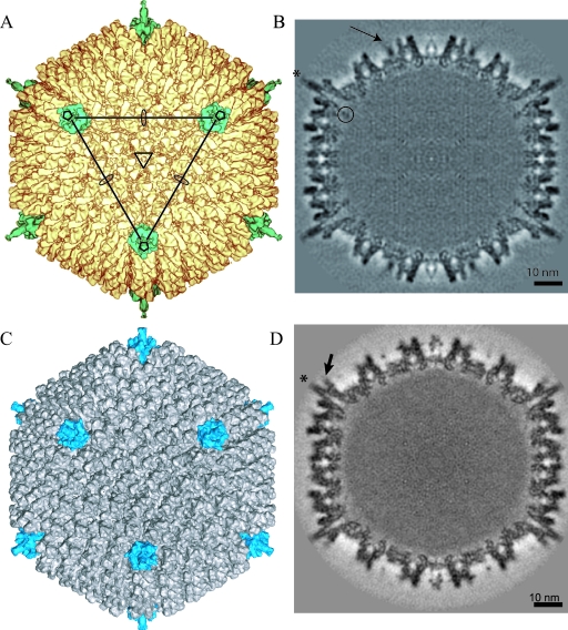
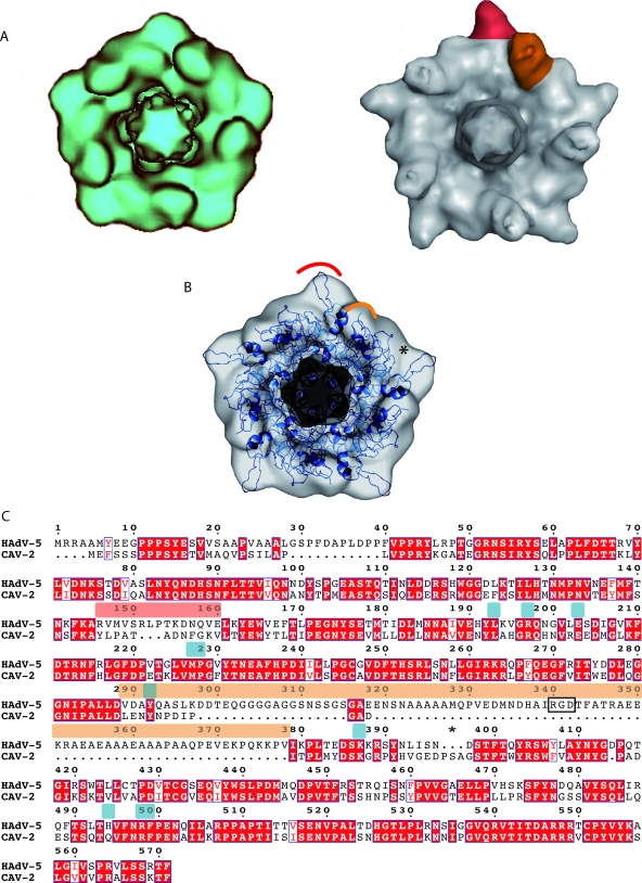
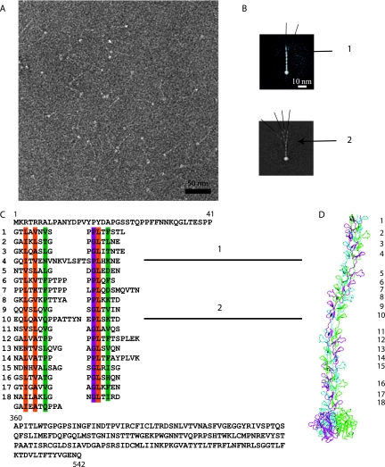
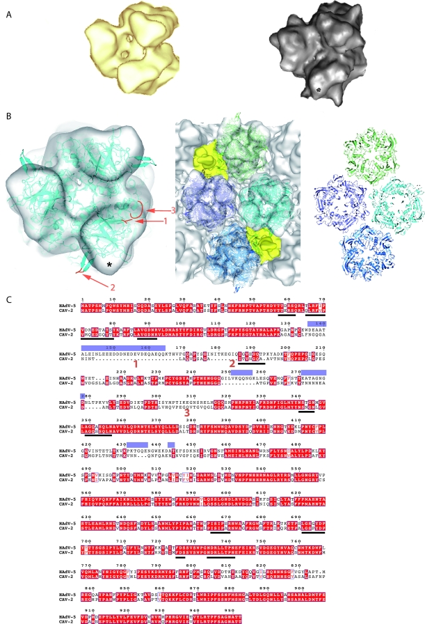
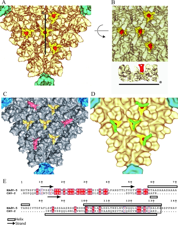
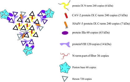
There are more than 100 known adenovirus (AdV) serotypes, including 50 human serotypes. Because AdV-induced disease is relatively species specific, vectors derived from nonhuman serotypes may have wider clinical potential based, in part, on the lack of ubiquitous memory immunity. Whereas a few of the human serotype capsids have been studied at the structural level, none of the nonhuman serotypes has been analyzed. The basis laid by the analysis of human AdV (hAdV) has allowed us to determine and compare the three-dimensional structure of the capsid of canine serotype 2 (CAV-2) to that of hAdV serotype 5 (hAdV-5). We show that CAV-2 capsid has a smoother structure than the human serotypes. Many of the external loops found in the hAdV-5 penton base and the hexon, against which the antibody response is directed, are shorter or absent in CAV-2. On the other hand, the CAV-2 fiber appears to be more complex, with two bends in the shaft. An interesting difference between the human and canine viruses is that the C-terminal part of protein IX is in a different position, making an antenna sticking out of the CAV-2 capsid. The comparison between the two viruses allows the identification of sites that should be easy to modify on the CAV-2 capsid for altering tissue tropism or other biological activities.


Figures

FIG. 1.
FIG. 1.
3D reconstruction of CAV-2 at 12-Å resolution and comparison with the previously determined structure of hAdV-5 (MSD accession number 1111) at 10-Å resolution. (A) Isosurface representation of CAV-2 as viewed down the threefold axis. The pentons are shown in petrol blue, and the rest of the capsid is shown in yellow. The icosahedral axes are indicated by an ellipse (twofold), a triangle (threefold), and a pentagon (fivefold). A continuous line connecting the three pentons pointing in your direction delimits a facet. (B) A central slice through the CAV-2 EM reconstruction showing that the capsid density is well defined. One of the fibers sticking out radially from the capsid is indicated by a star. A circle surrounds the nonattributed extra density present under the penton base. The arrow indicates protein IX density. Scale bar, 10 nm. (C) Isosurface representation of hAdV-5 view down the threefold axis. The pentons are shown in blue, and the rest of the capsid is shown in gray. (D) A central slice through the hAdV-5 EM reconstruction showing that the capsid density is well defined. The fibers sticking out radially from the capsid are indicated by a star. The RGD loop is indicated by an arrow. Scale bar, 10 nm.
FIG. 2.
FIG. 2.
The CAV-2 penton base. (A) Comparison between the computationally isolated penton from the CAV-2 capsid in blue (left) and the hAdV-5 capsid in gray (right). One of the five hypervariable regions is highlighted in red, and one protuberance harboring the RGD sequence is shown in orange in the hAdV-5 penton. (B) Fitting of the hAdV-2 penton base X-ray structure in blue (PDB accession number 1X9T) into the 12-Å CAV-2 cryo-EM envelope. The red and orange parts correspond to the regions indicated in panel A. The star indicates a region where the X-ray structure did not fill the EM density. (C) Sequence alignment of the hAdV-5 and CAV-2 penton bases visualized with ESPript (23). The RGD and hypervariable loops are indicated by bars above the sequence in the same colors as used in panels A and B. The RGD sequence is boxed with a black rectangle. Conserved residues are in white text and highlighted in red; partially conserved residues are in red text. The star highlights three extra amino acids present in CAV-2 compared to hAdV-5 that could fill up the density indicated by the star in panel B. The amino acids involved in fiber binding for hAdV-5 are indicated by blue bars above the sequence.
FIG. 3.
FIG. 3.
The CAV-2 fiber. (A) Electron micrograph of negatively stained CAV-2 fibers. The thin shafts and their globular knobs are clearly visible. Scale bar, 50 nm. (B) Averaged image of fibers selected from micrographs like that shown in panel A. The fiber is generally straight but can exhibit two kinks as indicated by the numbers 1 and 2. The different lines show the angle that the N-terminal part makes with the rest of the shaft. (C) Sequence of the CAV-2 fiber shaft showing the repeat motifs that correspond to the location of the bends. The top part of the sequence is the N-terminal tail of the fiber, and the lower part is the C-terminal knob. The repeat residues involved in the hydrophobic core are in orange, the ones forming the peripheral hydrophobic patches are in green, and the conserved glycine or proline residues are highlighted in purple according to van Raaij et al. (54). (D) Hypothetical model of the CAV-2 fiber showing the predicted bends at repeats 4 and 10. The repeats are indicated with the same numbers as in the sequence shown in panel C.
FIG. 4.
FIG. 4.
The CAV-2 hexon capsomer. (A) Comparison of computer-isolated hexons from CAV-2 (yellow) and hAdV-5 (gray) imaged in isodensity surface view. (B) Fitting of hAdV-5 hexon X-ray structure (PDB 1P30) into the CAV-2 cryo-EM envelope. Either one hexon alone (left) or the four hexons surrounding a twofold axis (middle) have been fitted using SITUS and imaged at the same time in the EM density. On the right, only the bottom wall of the pseudo-atomic model of the four hexons is shown. In the hexon alone, three regions are indicated on the X-ray structure; they correspond to large differences in the sequence of CAV-2 compared to that of hAdV-5 (see below). The red loops and regions 1 to 3 in the single hexon on the left are discussed in the text and correspond to sequence elements 1 to 3 indicated in panel C. The star indicates a region in the EM density that is not filled by the density derived from the crystal structure, as discussed in the text. (C) Sequence alignment between the CAV-2 and hAdV-5 hexon bases. Conserved residues are in white text and highlighted in red; partially conserved residues are in red text. The loops not resolved in the X-ray structure are indicated by blue bars above the sequences. The amino acid sequences involved in the contacts between hexons as described by Fabry et al. (15) are underlined with a bold line. They are conserved between CAV-2 and hAdV-5. Regions 1, 2, and 3 from panel B are indicated.
FIG. 5.
FIG. 5.
The minor protein IX. (A) Detailed view of one facet of CAV-2 seen down the threefold axis of the virion. The pentons are located on the vertices of the triangle and are shown in petrol blue, and the hexons are shown in pale yellow as in Fig. 1. Additional densities present as four groups of trimeric triskelions assigned to be protein IX are yellow (N-terminal part) and red (C-terminal part). (B) The top part represents the center of the facet described as in panel A but slightly tilted to see the extension of the C terminal part of protein IX form its N terminal part. The lower part is a slice through the capsid along the line shown in panel A. The color codes are the same as in panel A. (C) Detailed view of one facet of hAdV-5 seen down the threefold axis of the virion. The pentons are located on the vertices of the triangle and are in blue, and the hexons are gray. Protein IX is shown in yellow, and the cylindrical density that we initially assigned to protein IIIa (15) but that was recently reassigned to the C-terminal part of protein IX (45) is in red. (D) Detailed view of the threefold axis of the CAVpIX-GFP reconstruction. The penton and the hexons are as described in panel A. Protein IX is in yellow (N-terminal part) and red (C-terminal part), and the extra density assigned to GFP is in green. (E) Sequence alignment of hAdV-5 and CAV-2 protein IX. Conserved residues are in white text and highlighted in red; partially conserved residues are in red text. The arrows and open bars represent predicted β-strands and α-helices, respectively, as given by the secondary structure prediction software (http://bioweb.pasteur.fr/seqanal/interfaces/predator-simple.html). The predicted coil-coiled is boxed into a rectangle.
FIG. 6.
FIG. 6.
Schematic view of one facet of the CAV-2 capsid seen from the outside of the capsid. The figure shows a summary of all structural data on the protein localization in the AdV capsid. The hexons highlighted in black belong to the group of nine whereas the violet ones are the peripentonal hexons (26). The different proteins occupy the same positions in the capsid (hAdV and the CAV-2) except for the C-terminal part of protein IX that can either be on the top of the N terminus of protein IX in the CAV-2 capsid or be near the twofold axis in the hAdV capsid. For the hAdV protein IX, a thin black line indicates the position of the linker between the N- and the C-terminal parts of protein IX as suggested by Saban et al. (45).

References

    1. Akli, S., C. Caillaud, E. Vigne, L. D. Stratford-Perricaudet, L. Poenaru, M. Perricaudet, A. Kahn, and M. R. Peschanski. 1993. Transfer of a foreign gene into the brain using adenovirus vectors. Nat. Genet. 3224-228. - PubMed
    1. Baker, A. H., J. H. McVey, S. N. Waddington, N. C. Di Paolo, and D. M. Shayakhmetov. 2007. The influence of blood on in vivo adenovirus bio-distribution and transduction. Mol. Ther. 151410-1416. - PubMed
    1. Baker, T. S., and R. H. Cheng. 1996. A model-based approach for determining orientations of biological macromolecules imaged by cryoelectron microscopy. J. Struct. Biol. 116120-130. - PubMed
    1. Bergelson, J. M., J. A. Cunningham, G. Droguett, E. A. Kurt-Jones, A. Krithivas, J. S. Hong, M. S. Horwitz, R. L. Crowell, and R. W. Finberg. 1997. Isolation of a common receptor for coxsackie B viruses and adenoviruses 2 and 5. Science 2751320-1323. - PubMed
    1. Both, G. W. 2002. Xenogenic adenoviruses, p. 447-479. In D. Curiel and J. Douglas (ed.), Adenoviral vectors for gene therapy. Academic Press, San Diego, CA.

Publication types

MeSH terms

Substances

LinkOut - more resources