. 2008 Apr;82(7):3181-91.
doi: 10.1128/JVI.01612-07. Epub 2008 Jan 23.

NPXY motifs in the beta1 integrin cytoplasmic tail are required for functional reovirus entry

Affiliations

NPXY motifs in the beta1 integrin cytoplasmic tail are required for functional reovirus entry

Melissa S Maginnis et al. J Virol. 2008 Apr.

Abstract

Reovirus cell entry is mediated by attachment to cell surface carbohydrate and junctional adhesion molecule A (JAM-A) and internalization by beta1 integrin. The beta1 integrin cytoplasmic tail contains two NPXY motifs, which function in recruitment of adaptor proteins and clathrin for endocytosis and serve as sorting signals for internalized cargo. As reovirus infection requires disassembly in the endocytic compartment, we investigated the role of the beta1 integrin NPXY motifs in reovirus internalization. In comparison to wild-type cells (beta1+/+ cells), reovirus infectivity was significantly reduced in cells expressing mutant beta1 integrin in which the NPXY motifs were altered to NPXF (beta1+/+Y783F/Y795F cells). However, reovirus displayed equivalent binding and internalization levels following adsorption to beta1+/+ cells and beta1+/+Y783F/Y795F cells, suggesting that the NPXY motifs are essential for transport of reovirus within the endocytic pathway. Reovirus entry into beta1+/+ cells was blocked by chlorpromazine, an inhibitor of clathrin-mediated endocytosis, while entry into beta1+/+Y783F/Y795F cells was unaffected. Furthermore, virus was distributed to morphologically distinct endocytic organelles in beta1+/+ and beta1+/+Y783F/Y795F cells, providing further evidence that the beta1 integrin NPXY motifs mediate sorting of reovirus in the endocytic pathway. Thus, NPXY motifs in the beta1 integrin cytoplasmic tail are required for functional reovirus entry, which indicates a key role for these sequences in endocytosis of a pathogenic virus.


Figures
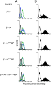
FIG. 1.
FIG. 1.
Reovirus exhibits equivalent binding to β1−/−, β1+/+, and β1+/+ cells with altered NPXY motifs. (A) GD25 (β1−/−), GD25β1A (β1+/+), GD25β1AY783F (β1+/+Y783F), GD25β1AY795F (β1+/+Y795F), and GD25β1AY783F/Y795F (β1+/+Y783F/Y795F) cells were detached from plates using 20 mM EDTA, washed, collected by centrifugation, and incubated with antibodies specific for either murine β1 integrin (blue), murine JAM-A (green), or an IgG control antibody (gray). Cell surface expression of these molecules was detected by flow cytometry. Data are expressed as fluorescence intensity. (B) β1−/−, β1+/+, β1+/+Y783F, β1+/+Y795F, and β1+/+Y783F/Y795F cells were detached from plates using 20 mM EDTA, washed, collected by centrifugation, and incubated with 104 particles per cell of reovirus T1L in incomplete medium (filled) or incomplete medium alone (open) at 4°C for 1 h to allow attachment. Cells were washed and incubated with reovirus-specific antiserum. Reovirus binding was detected by flow cytometry. Data are expressed as fluorescence intensity.
FIG. 2.
FIG. 2.
Reovirus infection is diminished in cells with altered β1 integrin NPXY motifs. β1−/−, β1+/+, β1+/+Y783F, β1+/+Y795F, and β1+/+Y783F/Y795F cells were adsorbed with T1L virions or ISVPs at an MOI of 1 FFU per cell at 4°C for 1 h. Cells were washed with PBS, incubated in complete medium at 37°C for 20 h, fixed, and stained by indirect immunofluorescence. Infected cells were quantified by counting cells exhibiting cytoplasmic staining in five visual fields of equivalently confluent monolayers for triplicate samples. The results are expressed as mean FFU per visual field for triplicate samples. Error bars indicate standard deviations. These data are representative of results of three individual experiments performed in triplicate. *, P < 0.05 in comparison to β1+/+ cells.
FIG. 3.
FIG. 3.
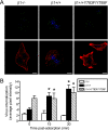
Reovirus is internalized into β1+/+ and β1+/+Y783F/Y795F cells. β1−/−, β1+/+, and β1+/+Y783F/Y795F cells were adsorbed with 5 × 10⁴ particles per cell of T1L virions and incubated at 4°C for 1 h. Nonadherent virus was removed, warm medium was added, and cells were incubated at 37°C. Cells were fixed over a time course, stained for reovirus (green), actin (red), and DNA (blue), and imaged using confocal immunofluorescence microscopy. (A) Representative digital fluorescence images of cells fixed at 30 min postadsorption are shown. Scale bars, 10 μm. (B) Fluorescent particles internalized into the cytoplasm were analyzed to determine the average pixel intensity of fluorescent particles per cell. Fluorescent particles localized at the cell periphery were excluded from the analysis. The results are expressed as the average pixel intensity per cell for five cells at each time point. Error bars indicate standard errors of the means. *, P < 0.05 in comparison to β1−/− cells.
FIG. 4.
FIG. 4.
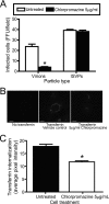
Uptake of reovirus and transferrin into β1+/+ cells is chlorpromazine sensitive. (A) β1+/+ cells were pretreated with 5 μg of chlorpromazine per ml for 3 h, adsorbed with T1L virions or ISVPs at an MOI of 1 FFU per cell, and incubated at 4°C for 1 h. Cells were washed, complete medium with or without chlorpromazine was added, and cells were incubated at 37°C for 20 h. Cells were fixed and stained by indirect immunofluorescence. Infected cells were quantified by counting cells exhibiting cytoplasmic staining in three visual fields of equivalently confluent monolayers for triplicate samples. The results are expressed as mean FFU per visual field for triplicate samples. Error bars indicate standard deviations. These data are representative of results of three independent experiments performed in triplicate. *, P < 0.05 in comparison to untreated cells. (B) β1+/+ cells were pretreated with 5 μg per ml of chlorpromazine for 3 h and incubated with 2.5 μg per ml Alexa Fluor 488-conjugated transferrin in the presence or absence of 5 μg per ml chlorpromazine in incomplete medium at 37°C for 10 min. The medium was removed, and cells were washed, fixed, and imaged by confocal microscopy. Representative digital fluorescence images of untreated and chlorpromazine-treated cells incubated with transferrin are shown. Scale bars, 10 μm. (C) Untreated and chlorpromazine-treated cells were analyzed for fluorescent transferrin internalized into the cytoplasm to determine the average pixel intensity of transferrin per cell. Fluorescent signal localized at the cell periphery was excluded from the analysis. The results are expressed as the average pixel intensity per cell for 10 cells. Error bars indicate standard errors of the means. *, P < 0.05 in comparison to untreated cells.
FIG. 5.
FIG. 5.
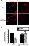
β1 integrin NPXY motifs are required for functional reovirus internalization. β1+/+ and β1+/+Y783F/Y795F cells were pretreated with 5 μg per ml of chlorpromazine at 37°C for 3 h, adsorbed with 5 × 10⁴ particles per cell of T1L virions in incomplete medium, and incubated at 4°C for 1 h. Cells where washed, complete medium with or without chlorpromazine was added, and cells were incubated at 37°C for 20 min. Cells were fixed, stained for reovirus (green), actin (red), and DNA (blue), and imaged using confocal immunofluorescence microscopy. (A) Representative digital fluorescence images of untreated and chlorpromazine-treated cells at 20 min postadsorption are shown. Scale bars, 10 μm. (B) Fluorescent particles internalized into the cytoplasm were analyzed to determine the average pixel intensity per cell. Fluorescent particles localized at the cell periphery were excluded from the analysis. The results are expressed as the average pixel intensity per cell for five cells. Error bars indicate standard errors of the means. *, P < 0.05 in comparison to untreated cells.
FIG. 6.
FIG. 6.
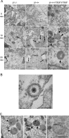
Ultrastructural analysis of reovirus internalization. (A) β1−/−, β1+/+, and β1+/+Y783F/Y795F cells were adsorbed with 105 particles per cell of T1L virions at 4°C for 1 h, washed, and fixed or incubated in complete medium at 37°C. At 10-min intervals, cells were washed, collected by centrifugation, fixed, and stained for EM. Representative images at 0, 10, and 20 min postadsorption are shown. Reovirus virions localized in coated pits and coated vesicles are indicated by white arrows. Reovirus virions localized in structures that resemble early and late endosomes and primary lysosomes are indicated by black arrows. Secondary lysosomes are indicated by black arrowheads. Scale bars, 500 nm. (B) Representative image of reovirus virion in a coated pit structure in β1+/+ cells at 0 min postadsorption. Scale bar, 100 nm. (C) Representative images of organelles resembling secondary lysosomes containing reovirus particles in β1+/+Y783F/Y795F cells at 10, 20, and 30 min postadsorption (left, center, and right, respectively). Reovirus virions are indicated by black arrows. Secondary lysosomes are indicated by black arrowheads. Scale bars, 500 nm.
FIG. 7.
FIG. 7.
Intracellular transport of reovirus particles is altered in β1+/+Y783F/Y795F cells. β1+/+ and β1+/+Y783F/Y795F cells were pretreated with Lysotracker-Red DND-99 dye (Lysotracker), adsorbed with 5 × 10⁴ particles per cell of T1L virions (Reovirus), and incubated at 4°C for 1 h. Nonadherent virus was removed, warm medium was added, and cells were incubated at 37°C. Cells were fixed over a time course and stained for reovirus. Images were analyzed for colocalization using ImageJ (version 1.37v) Colocalize RGB plug-in. Representative digital fluorescence images pseudocolored as monochromatic images or fluorescence images of cells fixed at 40 min postadsorption are shown. Lysotracker-Red DND-99 dye is colored red; reovirus is colored green. White arrowheads indicate areas of colocalization, which are magnified in the insets. Scale bars, 10 μm.

Similar articles

Cited by

References

    1. Agosto, M. A., T. Ivanovic, and M. L. Nibert. 2006. Mammalian reovirus, a nonfusogenic nonenveloped virus, forms size-selective pores in a model membrane. Proc. Natl. Acad. Sci. USA 10316496-16501. - PMC - PubMed
    1. Baer, G. S., and T. S. Dermody. 1997. Mutations in reovirus outer-capsid protein σ3 selected during persistent infections of L cells confer resistance to protease inhibitor E64. J. Virol. 714921-4928. - PMC - PubMed
    1. Bansal, A., and L. M. Gierasch. 1991. The NPXY internalization signal of the LDL receptor adopts a reverse-turn conformation. Cell 671195-1201. - PubMed
    1. Barton, E. S., J. L. Connolly, J. C. Forrest, J. D. Chappell, and T. S. Dermody. 2001. Utilization of sialic acid as a coreceptor enhances reovirus attachment by multistep adhesion strengthening. J. Biol. Chem. 2762200-2211. - PubMed
    1. Barton, E. S., J. C. Forrest, J. L. Connolly, J. D. Chappell, Y. Liu, F. Schnell, A. Nusrat, C. A. Parkos, and T. S. Dermody. 2001. Junction adhesion molecule is a receptor for reovirus. Cell 104441-451. - PubMed

Publication types