. 2008 Apr;82(7):3775-81.
doi: 10.1128/JVI.02154-07. Epub 2008 Jan 23.

Electron cryomicroscopy reveals different F1+F2 protein States in intact parainfluenza virions

Affiliations

Electron cryomicroscopy reveals different F1+F2 protein States in intact parainfluenza virions

Kai Ludwig et al. J Virol. 2008 Apr.

Abstract

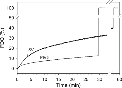
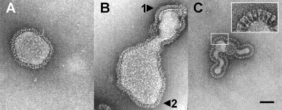
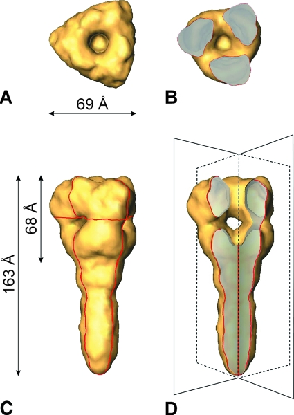
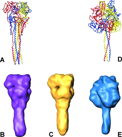
Electron cryomicrographs of intact parainfluenza virus 5 (PIV5) virions revealed two different surface structures, namely, a continuous layer and distinct individual spikes. The structure of these spikes reconstructed from intact virions was compared with known F ectodomain structures and was found to be different from the prefusion PIV5 F0 structure but, surprisingly, very similar to the human PIV3 F postfusion structure. Hence, we conclude that the individual F1+F2 spikes in intact PIV5 virions also correspond to the postfusion state. Since the observed fusion activity of PIV5 virions has to be associated with prefusion F1+F2 proteins, they have necessarily to be localized in the continuous surface structure. The data therefore strongly suggest that the prefusion state of the F1+F2 protein requires stabilization, most probably by the association with hemagglutinin-neuraminidase. The conversion of F1+F2 proteins from the prefusion toward the postfusion state while embedded in the virus membrane is topologically difficult to comprehend on the basis of established models and demands reconsideration of our current understanding.


Figures

FIG. 1.
FIG. 1.
Fusion kinetics of intact PIV5 and Sendai virus (SV) with human erythrocyte ghosts at 37°C and pH 7.4. R18-labeled PIV5 was bound to ghosts for 30 min at 4°C. Subsequently, fusion was initiated (0 min) by transfer of virosome cell complexes to prewarmed buffer (37°C). Fusion is measured by the release of self-quenching of R18 caused by redistribution of the fluorescent lipid analogue to the ghost plasma membrane. Fluorescence dequenching (FDQ) was normalized to the fluorescence intensity obtained after the addition of 0.5% Triton X-100 at the end of the kinetics (infinite dilution of the analogue).
FIG. 2.
FIG. 2.
Electron micrographs of a cryo-negative stain preparation of native PIV5 virions (strain W3A). Intact fusion-active virions were embedded in a matrix of vitreous ice in the presence of 1% (wt/vol) phosphotungstic acid. The surface structure of the envelopes is heterogeneous: either a continuous layer with no identifiable ultrastructure (A and B, arrow 1) or distinct individual spikes (B, arrow 2, and C) are detectable. Bar, 500 Å.
FIG. 3.
FIG. 3.
Surface presentation of the 3D reconstruction of the ectodomain of the PIV5 F protein, determined from cryo-negative stain electron micrographs at a resolution of ∼15 Å. (A) Top view. (C) Side view. The viral membrane (not shown) is located at the lower end of the stalk region. (B) Top view of a horizontal section (the localization of the cross section is indicated by the horizontal red line in panel C). (D) Axial cutaway view (45°) of panel C to reveal the axial channel and the three radial channels merging into the central cavity. The imaginary cutting planes are drawn in fine black lines (compare to the red contours in panel C).
FIG. 4.
FIG. 4.
Comparison of X-ray and electron cryomicroscopy structures of paramyxovirus F proteins. (A and B) hPIV3 “postfusion” X-ray structure (Protein Data Bank entry 1ZTM), Gaussian filtered at 15-Å resolution (surface presentation is shown in purple). (C) PIV5 in situ structure at 15-Å resolution, reconstructed from electron cryomicroscopy data. (D and E) PIV5 “prefusion” X-ray structure (Protein Data Bank entry 2B9B), Gaussian filtered at 15-Å resolution (blue).

References

    1. Baker, K. A., R. E. Dutch, R. A. Lamb, and T. S. Jardetzky. 1999. Structural basis for paramyxovirus-mediated membrane fusion. Mol. Cell 3309-319. - PubMed
    1. Beniac, D. R., A. Andonov, E. Grudeski, and T. F. Booth. 2006. Architecture of the SARS coronavirus prefusion spike. Nat. Struct. Mol. Biol. 13751-752. - PMC - PubMed
    1. Blumenthal, R., A. Bali-Puri, A. Walter, D. Covell, and O. Eidelman. 1987. pH-dependent fusion of vesicular stomatitis virus with Vero cells. Measurement by dequenching of octadecyl rhodamine fluorescence. J. Biol. Chem. 26213614-13619. - PubMed
    1. Böttcher, C., K. Ludwig, A. Herrmann, M. van Heel, and H. Stark. 1999. Structure of influenza hemagglutinin at neutral and at fusogenic pH by electron cryo-microscopy. FEBS Lett. 463255-259. - PubMed
    1. Chan, D. C., and P. S. Kim. 1998. HIV entry and its inhibition. Cell 93681-684. - PubMed

Publication types

LinkOut - more resources