Comparative Study
. 2008 Jan 23;28(4):923-31.
doi: 10.1523/JNEUROSCI.4012-07.2008.

Does it look painful or disgusting? Ask your parietal and cingulate cortex

Affiliations
Comparative Study

Does it look painful or disgusting? Ask your parietal and cingulate cortex

Francesca Benuzzi et al. J Neurosci. .

Abstract

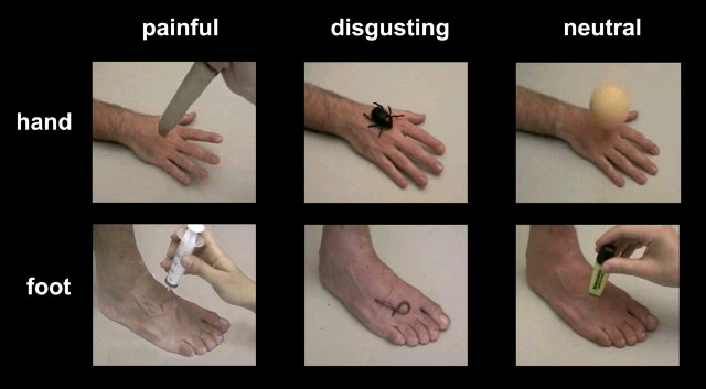
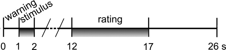
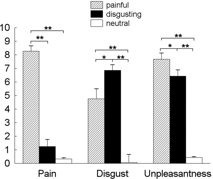
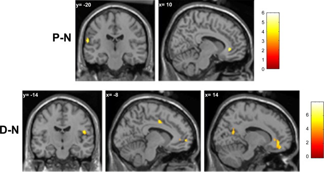
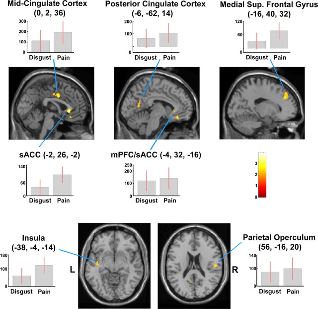
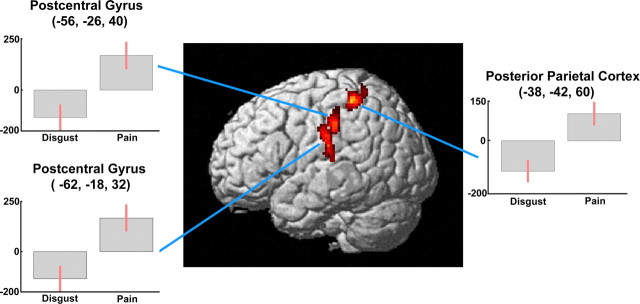
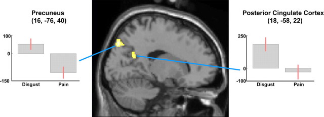
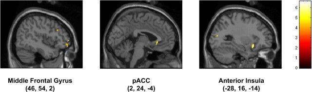
Looking at still images of body parts in situations that are likely to cause pain has been shown to be associated with activation in some brain areas involved in pain processing. Because pain involves both sensory components and negative affect, it is of interest to explore whether the visually evoked representations of pain and of other negative emotions overlap. By means of event-related functional magnetic resonance imaging, here we compare the brain areas recruited, in female volunteers, by the observation of painful, disgusting, or neutral stimuli delivered to one hand or foot. Several cortical foci were activated by the observation of both painful and disgusting video clips, including portions of the medial prefrontal cortex, anterior, mid-, and posterior cingulate cortex, left posterior insula, and right parietal operculum. Signal changes in perigenual cingulate and left anterior insula were linearly related to the perceived unpleasantness, when the individual differences in susceptibility to aversive stimuli were taken into account. Painful scenes selectively induced activation of left parietal foci, including the parietal operculum, the postcentral gyrus, and adjacent portions of the posterior parietal cortex. In contrast, brain foci specific for disgusting scenes were found in the posterior cingulate cortex. These data show both similarities and differences between the brain patterns of activity related to the observation of noxious or disgusting stimuli. Namely, the parietal cortex appears to be particularly involved in the recognition of noxious environmental stimuli, suggesting that areas involved in sensory aspects of pain are specifically triggered by observing noxious events.


Figures

Figure 1.
Figure 1.
Sample frames extracted from some video clips representing painful (left), disgusting (middle), and neutral (right) stimuli. All video clips began with 200–400 ms of a static hand or foot picture, followed by a stimulus rapidly approaching and contacting the skin. Needles and knifes apparently punctured the hand or foot, but actually they did not: the images were digitally corrected to simulate bleeding.
Figure 2.
Figure 2.
Experimental design. Temporal sequence of events during each trial. For details, see Materials and Methods.
Figure 3.
Figure 3.
On-line (unpleasantness) and off-line (disgust and painfulness) ratings of visually presented stimuli. *p < 0.05; **p < 0.0001.
Figure 4.
Figure 4.
Top, Cortical foci active during the observation of painful video clips, identified from the (P − N) contrast. Bottom, Cortical foci active during the observation of disgusting video clips, identified from the (D − N) contrast. Maps are thresholded at p < 0.001 uncorrected for multiple comparisons, with a cluster extent threshold (k) of 25 voxels in the interpolated images (200 mm3, p < 0.05).
Figure 5.
Figure 5.
Clusters showing higher activity during the observation of both painful and disgusting versus neutral video clips [(P − N) + (D − N)]. Cingulate and prefrontal foci are displayed on sagittal sections (top), left insular and right parietal foci on axial sections (bottom) from the MNI template. Plots of parameter estimates (mean ± SEM of 15 subjects) of the effects of P − N and D − N observation for peak activity voxels are also shown. Random-effect analysis, height threshold of p < 0.001 and cluster threshold of p < 0.05 (both uncorrected). R, Right side of the brain; L, left side of the brain.
Figure 6.
Figure 6.
Three-dimensional rendering showing cortical clusters in the left parietal cortex, more active during the observation of painful than disgusting video clips [(P − N) − (D − N)]. Plots of parameter estimates for peak activity voxels refer to the P − N and D − N contrasts, respectively.
Figure 7.
Figure 7.
Sagittal section showing cortical clusters more active during the observation of disgusting than painful video clips [(D − N) − (P − N)]. Plots of parameter estimates for peak activity voxels refer to the P − N and D − N contrasts, respectively.
Figure 8.
Figure 8.
Sagittal sections showing cortical clusters whose activity was linearly correlated with the perceived unpleasantness and susceptibility to fearful stimuli (for details, see Materials and Methods).

References

    1. Adolphs R. Neural systems for recognizing emotion. Curr Opin Neurobiol. 2002;12:169–177. - PubMed
    1. Adolphs R, Damasio H, Tranel D, Cooper G, Damasio AR. A role for somatosensory cortices in the visual recognition of emotion as revealed by three-dimensional lesion mapping. J Neurosci. 2000;20:2683–2690. - PMC - PubMed
    1. Albanese MC, Duerden EG, Rainville P, Duncan GH. Memory traces of pain in human cortex. J Neurosci. 2007;27:4612–4620. - PMC - PubMed
    1. Avenanti A, Bueti D, Galati G, Aglioti SM. Transcranial magnetic stimulation highlights the sensorimotor side of empathy for pain. Nat Neurosci. 2005;8:955–960. - PubMed
    1. Avenanti A, Paluello IM, Bufalari I, Aglioti SM. Stimulus-driven modulation of motor-evoked potentials during observation of others' pain. NeuroImage. 2006;32:316–324. - PubMed

Publication types