Comparative Study
. 2008 Jan 23;28(4):944-52.
doi: 10.1523/JNEUROSCI.2934-07.2008.

Parallel processing of nociceptive A-delta inputs in SII and midcingulate cortex in humans

Affiliations
Comparative Study

Parallel processing of nociceptive A-delta inputs in SII and midcingulate cortex in humans

Maud Frot et al. J Neurosci. .

Abstract

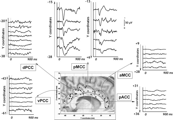
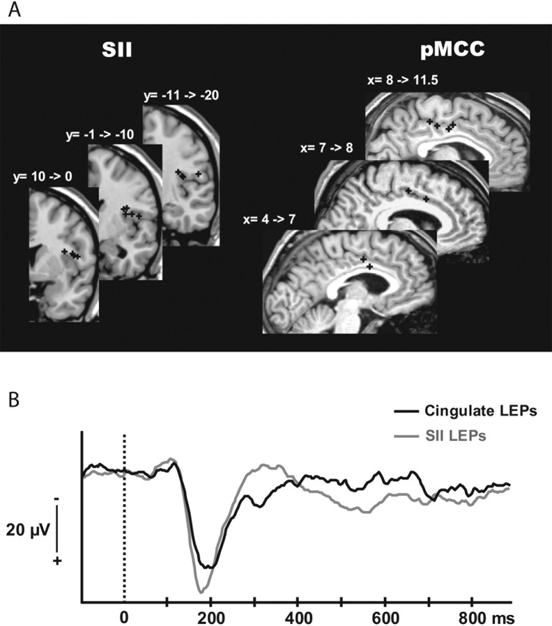
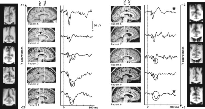
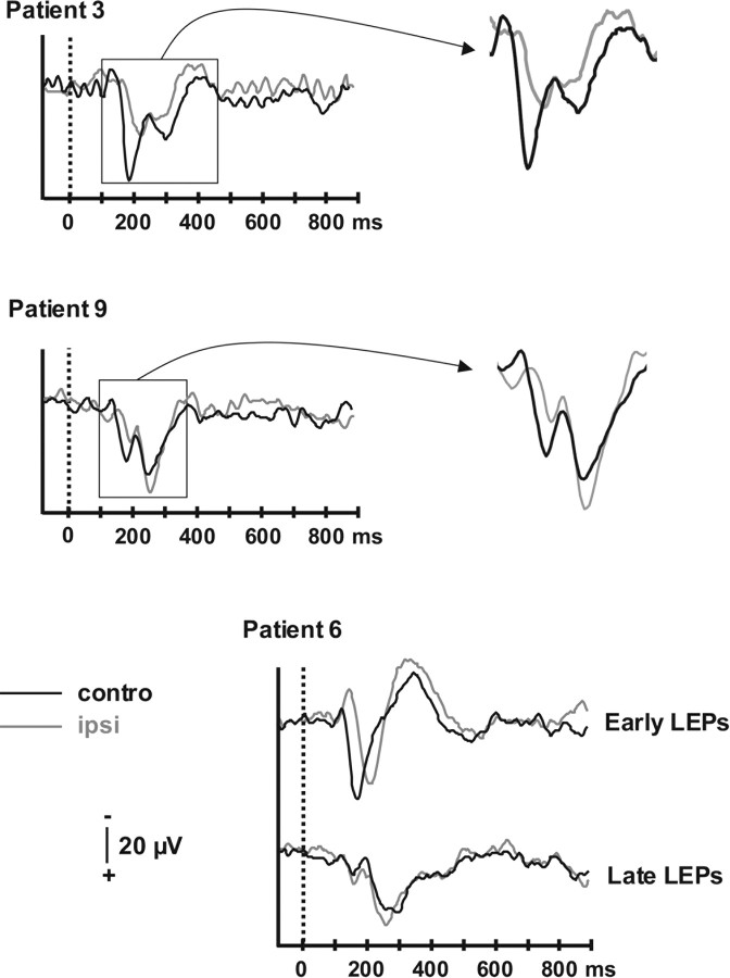
The cingulate cortex (CC) as a part of the "medial" pain subsystem is generally assumed to be involved in the affective and/or cognitive dimensions of pain processing, which are viewed as relatively slow processes compared with the sensory-discriminative pain coding by the lateral second somatosensory area (SII)-insular cortex. The present study aimed at characterizing the location and timing of the CC evoked responses during the 1 s period after a painful laser stimulus, by exploring the whole rostrocaudal extent of this cortical area using intracortical recordings in humans. Only a restricted area in the median CC region responded to painful stimulation, namely the posterior midcingulate cortex (pMCC), the location of which is consistent with the so-called "motor CC" in monkeys. Cingulate pain responses showed two components, of which the earliest peaked at latencies similar to those obtained in SII. These data provide direct evidence that activations underlying the processing of nociceptive information can occur simultaneously in the "medial" and "lateral" subsystems. The existence of short-latency pMCC responses to pain further indicates that the "medial pain system" is not devoted exclusively to the processing of emotional information, but is also involved in fast attentional orienting and motor withdrawal responses to pain inputs. These functions are, not surprisingly, conducted in parallel with pain intensity coding and stimulus localization specifically subserved by the sensory-discriminative "lateral" pain system.


Figures

Figure 1.
Figure 1.
CC recordings in all patients. Explored cingulate sites pooled across patients (bipolar montage). All contacts were plotted in the stereotactic system of Talairach and Tournoux and on one MRI. This parasagittal MRI was chosen in the pool of the MRI of all patients included in this study; therefore, some of the sites seem to be located outside the limits of the CC. However, they were truly situated in the CC when plotted on the appropriate parasagittal slices in each individual (see Fig. 3 for pMCC). Recording sites were clustered in five of the six subregions individualized by Vogt (2005): pACC, aMCC, pMCC, dPCC, and vPCC.
Figure 2.
Figure 2.
SII versus cingulate LEPs. A, Location of the SII and pMCC contacts where the maximal amplitudes of the N/P deflection in bipolar mode were recorded. The contacts were plotted on MRI slices chosen in the pool of the MRI of all patients included in the study, according to their Talairach coordinates. The precise SII and pMCC location of all these contacts was verified by plotting them on the appropriate MRI slices of each patient. B, Grand average LEPs in bipolar recording mode from all the patients in SII (gray) and CC (black).
Figure 3.
Figure 3.
LEPs recorded in the pMCC and location of the contacts. Evoked responses recorded in the pMCC cluster (see Fig. 1) in nine patients. The location of the contacts (black dots and black crosses) presenting such responses were plotted on sagittal and coronal MRI slices of each patient. The more posterior location is situated in the bottom left corner of the figure, and the more anterior location is situated in the bottom right corner. An early CC LEP component was recorded in all patients (black arrows) on contacts located between vertical planes +6 mm anterior and −28 mm posterior to the VAC coronal plane (y coordinates). We also recorded a late CC LEP component (in black circles) in six patients on the more posterior (−28 mm < y < −20 mm) and more anterior (−2 mm < y < +6 mm) contacts within the pMCC, but not on the contacts located between −18 and −9 mm from the VAC coronal plane. In one patient, early and late components of the CC LEPs were separately recorded on two distinct electrode tracks (patient 6, black star). VPC, Vertical posterior commissure plane
Figure 4.
Figure 4.
Contralateral versus ipsilateral CC LEPs. Cortical cingulate responses evoked by YAP laser stimuli recorded contralateral (black traces) and ipsilateral (gray traces) to the stimulated hand in three patients. Early and late components of CC LEPs were recorded on the same contacts for patients 3 and 9 and on two different contacts for patient 6. Early ipsilateral LEPs peak ∼17 ms later than their contralateral homologues, whereas there was no latency difference between contralateral and ipsilateral late CC LEPs.

References

    1. Adriaensen H, Gybels J, Handwerker HO, Van Hees J. Response properties of thin myelinated (A-delta) fibers in human skin nerves. J Neurophysiol. 1983;49:111–122. - PubMed
    1. Albe-Fessard D, Berkley KJ, Kruger L, Ralston HJ, III, Willis WD., Jr Diencephalic mechanisms of pain sensation. Brain Res. 1985;356:217–296. - PubMed
    1. Apkarian VA, Hodge CJ. Primate spinothalamic pathways: I. A quantitative study of the cells of origin of the spinothalamic pathway. J Comp Neurol. 1989;288:447–473. - PubMed
    1. Baleydier C, Mauguière F. The duality of the cingulate gyrus in monkey—neuroanatomical study and functional hypothesis. Brain. 1980;103:525–554. - PubMed
    1. Bastuji H, Perchet C, Legrain V, Montes C, Garcia-Larrea L. Laser evoked responses to painful stimulation persist during sleep and predict subsequent arousals. Pain. 2008 in press. - PubMed

Publication types