Comparative Study
. 2008 Jan 23;28(4):953-62.
doi: 10.1523/JNEUROSCI.3237-07.2008.

Exercise-induced activation of NMDA receptor promotes motor unit development and survival in a type 2 spinal muscular atrophy model mouse

Affiliations
Comparative Study

Exercise-induced activation of NMDA receptor promotes motor unit development and survival in a type 2 spinal muscular atrophy model mouse

Olivier Biondi et al. J Neurosci. .

Abstract

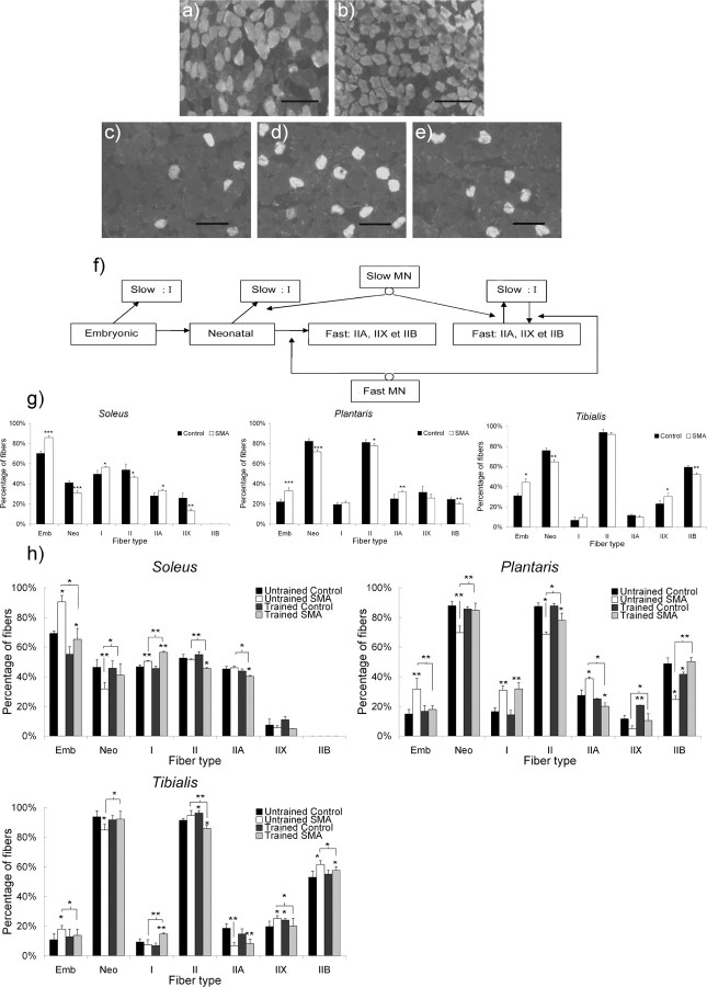
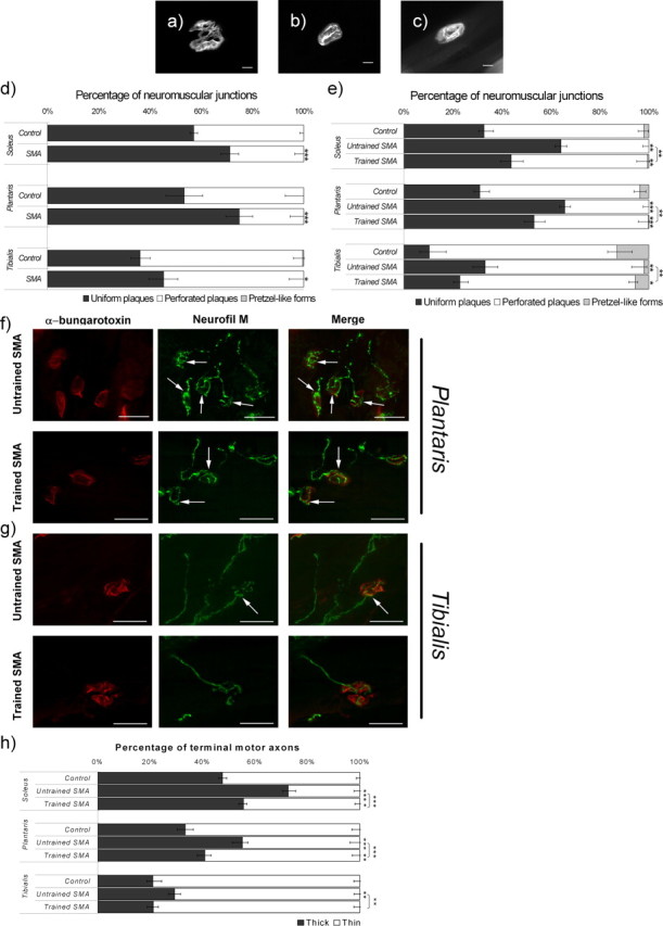
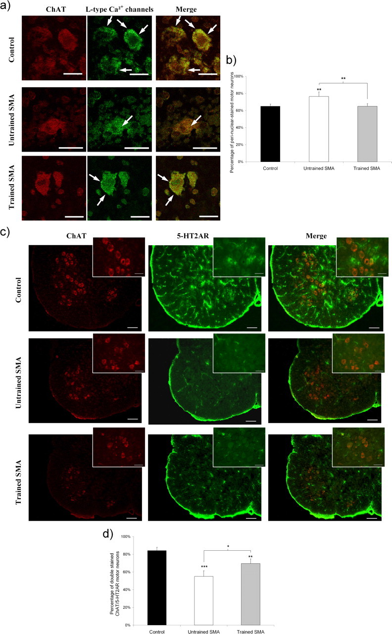
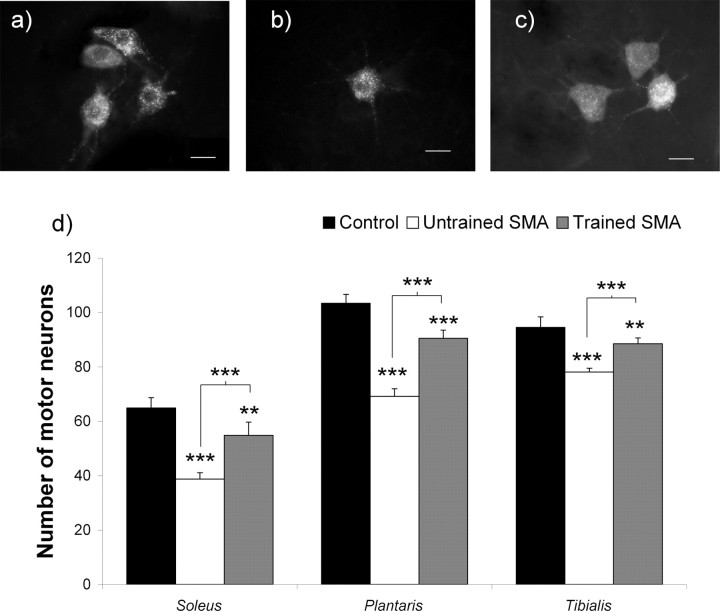
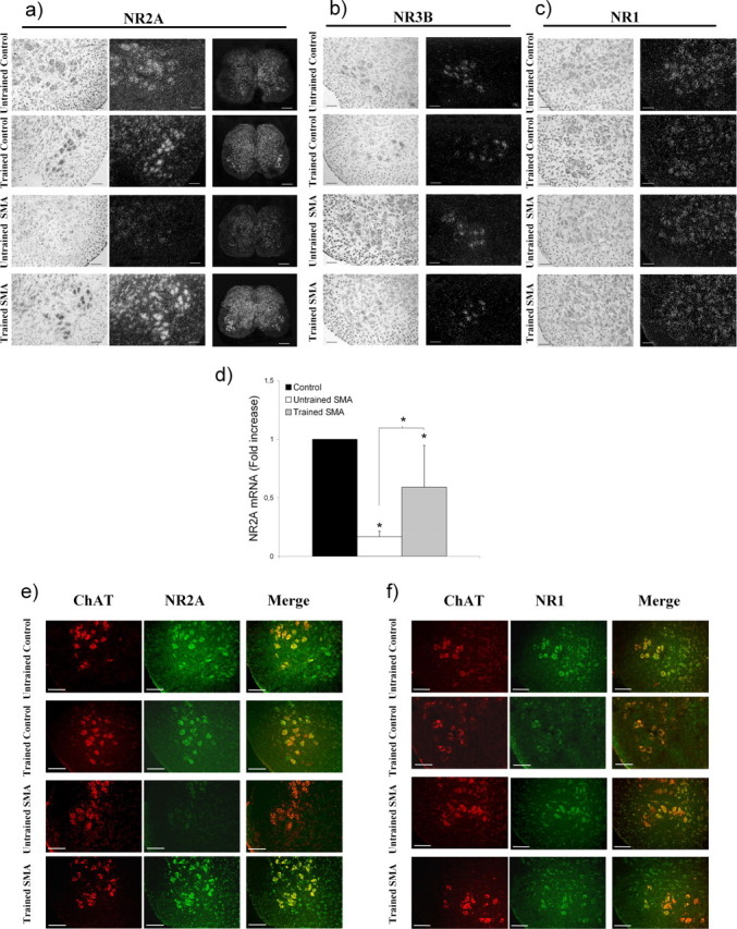
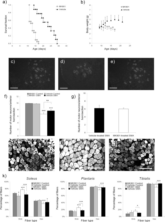
Spinal muscular atrophy (SMA) is an inborn neuromuscular disorder caused by low levels of survival motor neuron protein, and for which no efficient therapy exists. Here, we show that the slower rate of postnatal motor-unit maturation observed in type 2 SMA-like mice is correlated with the motor neuron death. Physical exercise delays motor neuron death and leads to an increase in the postnatal maturation rate of the motor-units. Furthermore, exercise is capable of specifically enhancing the expression of the gene encoding the major activating subunit of the NMDA receptor in motor neurons, namely the NR2A subunit, which is dramatically downregulated in the spinal cord of type 2 SMA-like mice. Accordingly, inhibiting NMDA-receptor activity abolishes the exercise-induced effects on muscle development, motor neuron protection and life span gain. Thus, restoring NMDA-receptor function could be a promising therapeutic approach to SMA treatment.


Figures

Figure 1.
Figure 1.
Muscle typology analysis of the extensor slow-twitch soleus, the extensor fast-twitch plantaris, and the flexor fast-twitch tibialis from type 2 SMA-like mice. a–e, Immunodetection of MyHC isoforms revealed an increase in embryonic muscle fiber proportion in the soleus from type 2 SMA-like mice (b) compared with control mice (a) at 9 d of age and an increase in type I muscle fiber proportion in the plantaris from untrained SMA-like mice (d) compared with trained SMA-like mice (e) and control mice (c) at 12 d of age. Scale bars, 100 μm. f, Schematic presentation of the myofiber transitions during postnatal development of the motor unit including the role of motor neuron innervations on adult phenotype acquisition. MN, Motor neuron; Emb, embryonic; Neo, neonatal. g, h, The quantitative analysis of the muscle typology variation during the postnatal development of soleus, plantaris, and tibialis muscles from type 2 SMA-like mice compared with control at 9 d of age (g) and from untrained and trained type 2 SMA-like mice compared with controls at 12 d of age (h) shows a muscle-function-specific delay in skeletal-muscle maturation in SMA that could be counteracted by the exercise protocol (n = 6; *p < 0.05; **p < 0.01; ***p < 0.001).
Figure 2.
Figure 2.
Morphological analysis of the NMJs in the soleus, plantaris, and tibialis from type 2 SMA-like mice. a–c, Uniform immature plaque of NMJ are frequently observed in tibialis from untrained SMA-like mice (b) compared with the more perforated plaque, which is preferentially observed in trained SMA-like mice (c) and the pretzel-like form observed in control mice at 12 d of age (a). Scale bars, 10 μm. d, e, Changes in the NMJ morphology computed after a whole-mount α-bungarotoxin labeling of soleus, plantaris, and tibialis muscles from untrained and trained type 2 SMA-like mice compared with control mice at 9 d (d) and 12 d of age (e) reveal an NMJ maturation delay in SMA that could be counteracted by the exercise protocol. f, AChR (red) and neurofilament (Neurofil M) (green) labeling in the plantaris muscle reveal the presence of thick terminal axons (arrows) at the NMJ of untrained (top) type 2 SMA-like mice, whereas thin terminal axons are more frequently observed in trained type 2 SMA-like mice (bottom) at 12 d of age. g, These morphological differences are less evident in the tibialis muscle. Scale bars, 25 μm. h, Quantification of thick terminal axons filled with neurofilament in the soleus, plantaris, and tibialis muscles from untrained and trained type 2 SMA-like mice and from control mice (n = 6; *p < 0.05; **p < 0.01; ***p < 0.001).
Figure 3.
Figure 3.
Analysis of motor neuron postnatal maturation in untrained and trained type 2 SMA-like mice. a, The subcellular location of the L-type calcium channels as determined by confocal immunofluorescence imaging is preferentially a perinuclear one (arrows), in the motor neurons of the lumbar spinal cord from untrained type 2 SMA-like mice (middle) compared with membrane-labeled motor neurons (arrows) from trained SMA-like mice (bottom) and control mice (top) at 12 d of age. Scale bars, 10 μm. b, The proportion of perinuclear-stained motor neurons recorded in the spinal cord of untrained SMA-like mice is significantly decreased in the spinal cord of trained SMA-like and control mice at 12 d of age (n = 6, **p < 0.01). c, The motor neuron maturation rate was further evaluated by the immunodetection level of the 5-HT2AR over the lumbar spinal cord of the untrained (middle) versus trained (bottom) SMA-like mice and control mice (top). Scale bars: low-magnification views, 100 μm; high-magnification views, 25 μm. d, The quantification of 5-HT2AR-expressing motor neurons highlights a delay in 5-HT2AR appearance in the lumbar spinal cord from untrained type 2 SMA-like mice when compared with their trained counterparts (n = 4; *p < 0.05; **p < 0.01; ***p < 0.001).
Figure 4.
Figure 4.
Analysis of the muscle-specific motor-neuron populations in untrained and trained type 2 SMA-like mice. a–c, A loss of the soleus-innervating motor neurons, specifically labeled using a fluorogold retrolabeling technique, is observed in the spinal cord from untrained type 2 SMA-like mice (b) compared with trained type 2 SMA-like (c) and control mice (a) at 12 d of age. Scale bars, 20 μm. d, The quantification of surviving motor neurons innervating each muscle clearly indicates that the extent of motor neuron loss in type 2 SMA-like mice at 12 d of age is muscle-specific and that, in each muscle, the exercise protocol exerts an efficient neuroprotective effect in SMA (n = 6; *p < 0.05; **p < 0.01; ***p < 0.001).
Figure 5.
Figure 5.
Expression profiles of NMDA-receptor isoforms in the lumbar spinal cord from trained and untrained type 2 SMA-like mice. a–c, An important decrease in the NR2A subunit expression level is observed in the lumbar spinal cord of the type 2 SMA-like mice at 12 d of age at the level of the transcripts compared with controls (a) whereas the expression of either the NR3B (b) or the NR1 (c) subunit genes remains unchanged. The exercise regimen proves to specifically enhance the NR2A subunit gene expression in SMA-like as well as in control mice. Scale bars: 50 μm for all images, except 250 μm for the large views of the spinal cord. d, The quantification of NR2A transcripts in the spinal cord at 12 d of age by real-time RT-PCR further substantiates the assumption of an exercise-induced reactivation of the NR2A gene expression in trained SMA-like mice (n = 3; *p < 0.05). e, The immunodetection level of NR2A in motor neurons, identified for their immunoreactivity for ChAT, is in agreement with the in situ hybridization data. The low NR2A labeling in motor neurons from untrained type 2 SMA-like mice is to be compared with the high detection of this protein in the motor neurons of trained SMA-like mice, at 12 d of age. Scale bars, 50 μm. f, In contrast, no difference in the NR1 distribution in the spinal cord of SMA-like and control mice could be evidenced (n = 4).
Figure 6.
Figure 6.
NMDA-receptor involvement in the exercise-induced neuroprotection in type 2 SMA-like mice. a–e, SMA-like mice that were submitted to a MK-801 treatment (MK801; n = 21) before training have lost the benefits of the exercise regimen compared with the vehicle-treated trained mice (Vehicle; n = 9) in terms of life span (12.8 and 22.3 d, respectively), as revealed by the Kaplan-Meier curve (a); growth gain, as revealed by the growth curve (b); and motor neuron protection, as revealed by ChAT-immunodetection (c–e). Scale bars, 50 μm. f, The quantification of the motor neurons was performed in the anterior horn of the lumbar spinal cord (L1–L5) from the MK-801- or vehicle-treated trained SMA-like mice (n = 7 and n = 6, respectively) and compared with MK-801- or vehicle-treated trained control mice (n = 6) (*p < 0.05; **p < 0.01; ***p < 0.001). g, The MK-801 treatment does not promote motor neuron death at 12 d of age in the spinal cord of sedentary type 2 SMA-like mice, treated by MK-801 or vehicle, as revealed by the count of Chat-positive cells (n = 3; nonsignificant). h, j, Immunodetection of MyHC isoforms revealed an increase in embryonic myofiber proportion in the soleus from MK-801-treated (i) versus vehicle-treated (j) trained type 2 SMA-like mice and control mice (h) at 12 d of age. Scale bars, 100 μm. k, The comparison of the muscle typology of the soleus, plantaris, and tibialis muscles in MK-801- versus vehicle-treated trained type 2 SMA-like and control mice at 12 d of age (n = 8, 6, 6, and 6, respectively) indicate that the NMDA-receptor blockade results in the abolishment of the exercise-induced acceleration of muscle maturation (*p < 0.05; **p < 0.01; ***p < 0.001).

References

    1. Abercrombie M. Estimation of nuclear population from microtome sections. Anat Rec. 1946;94:239–247. - PubMed
    1. Andersson O, Stenqvist A, Attersand A, von Euler G. Nucleotide sequence, genomic organization, and chromosomal localization of genes encoding the human NMDA receptor subunits NR3A and NR3B. Genomics. 2001;78:178–184. - PubMed
    1. Andreassi C, Patrizi AL, Monani UR, Burghes AH, Brahe C, Eboli ML. Expression of the survival of motor neuron (SMN) gene in primary neurons and increase in SMN levels by activation of the N-methyl-d-aspartate glutamate receptor. Neurogenetics. 2002;4:29–36. - PubMed
    1. Brichta L, Hofmann Y, Hahnen E, Siebzehnrubl FA, Raschke H, Blumcke I, Eyupoglu IY, Wirth B. Valproic acid increases the SMN2 protein level: a well known drug as a potential therapy for spinal muscular atrophy. Hum Mol Genet. 2003;12:2481–2489. - PubMed
    1. Dick J, Greensmith L, Vrbova G. Blocking of NMDA receptors during a critical stage of development reduces the effects of nerve injury at birth on muscles and motoneurones. Neuromuscul Disord. 1995;5:371–382. - PubMed

Publication types

MeSH terms

Substances

LinkOut - more resources